Interesting findings
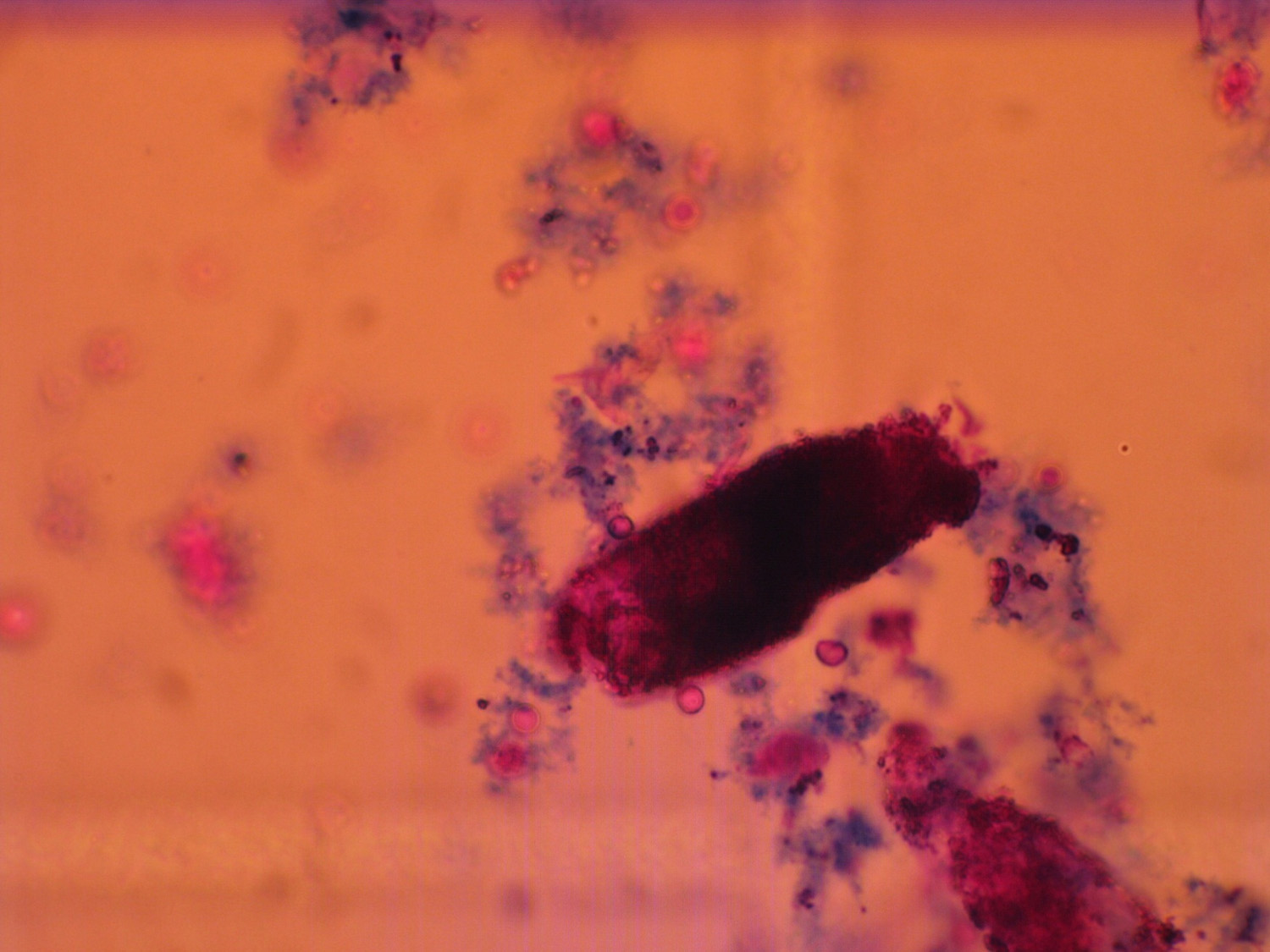
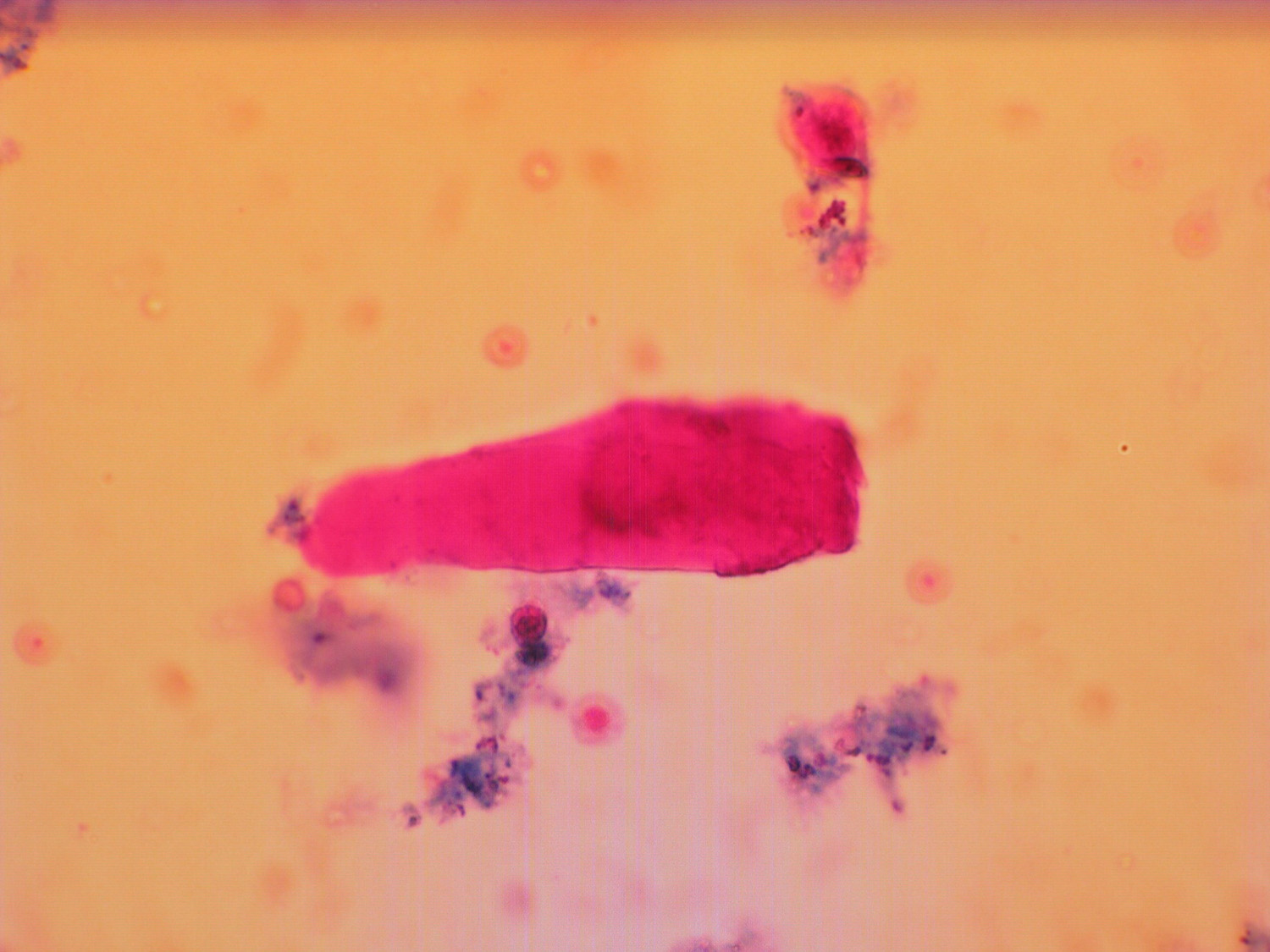
Erythrocytes x yeast
Occasionally, it is difficult to distinguish between single yeast and erythrocyte due to their similar shape and size in the pictures from automatic analyzer and in native sediment. The stained sediment can be used for determinative analysis, because erythrocytes are typically stained pink, but yeasts are colorless.

Yeast between erytrocytes
Erythrocytes and yeast together
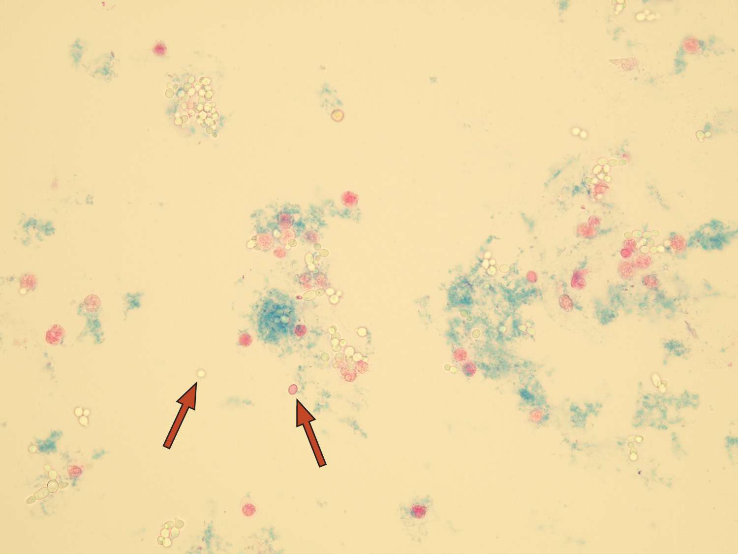
Erythrocytes x oxalates
If erythrocytes and oxalates are both in urine, it is sometimes difficult to distinguish them in the automatic analyzer properly due to their similar size and shape. This applies particularly to calcium oxalate monohydrate (ovoid form). In the stained sediment on the other hand, the erythrocytes are pink and oxalates colorless.

Calcium oxalate monohydrate crystals next to erythrocytes automatically classified as erythrocytes in FUS-2000

Calcium oxalate monohydrate crystals next to erythrocytes
The gradual transformation of pathological casts
Exceptionally, all stages of cast transformations could be observed in one sample.

Cellular cast

Transition phase of cast from cellular to granular (enlargement ×600)

Transition phase of cast from cellular to granular
Granular cast

Transition phase of cast from granular to waxy and waxy cast
Waxy cast
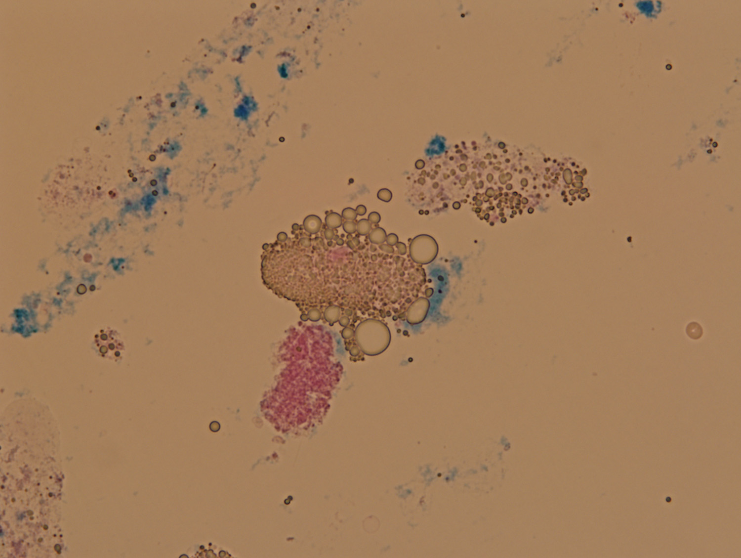
The finding of fat particles in patient with nephrotic syndrome
The elements listed below were found in urine of patient with nephrotic syndrome.

Hyaline cast with fat inclusions of fat droplets

Squamous epithelial cell and oval fat body
Fatty cast and free fat droplets
Elements in hypotonic urine
A patient with acute urine bladder inflammation was treated by hydration of the bladder, causing his urine to be hypotonic.
In the urine sample, we found neutrophil granulocytes which are called “glitter cells” because of the rapid Brownian movement of granules inside their cells. The low urine osmolality caused ruptures in the cell membranes of several of these leukocytes which resulted in cytoplasm spilling outside of the cells. These cells are sometimes called “winged leukocytes”.
The hypotonic urine from the same patient also caused erythrocytes to swell and their size matched that of the leukocytes. The same size resulted in miscategorization of some erythrocytes in the leukocyte category by the FUS-2000 analyzer software leading to a discrepant finding.

Leukocytes – some of them glittering cells (pointer)

Swollen erythrocytes in hypotonic urine
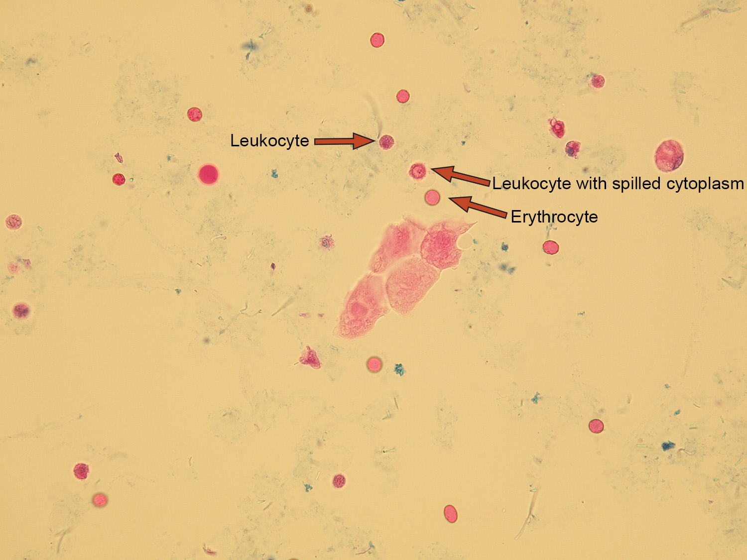
Leukocytes (glittering cells) with erythrocytes

Leukocytes (glittering cells) with erythrocytes
Sample contaminated by feces
The urine analyzed with FUS-2000 showed some dark asymmetric particles miscategorized as squamous epithelial cells. Further investigation with microscopy in stained and native sediment confirmed them to be stool particles. This rare contamination of feces may occur in patients with fistula of bladder.

Fecal particles (black elements) miscategorized in squamous epithelia section
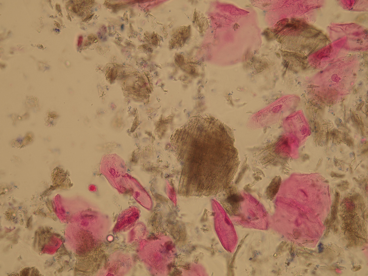
Fecal particles (brown elements) with squamous cells
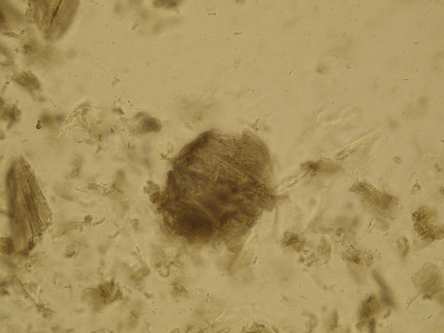
Fecal particles
Intracellular Bacteria
Bacteria, most commonly E. coli, may be sometimes observed inside epithelial cells of the bladder in samples of patients with urinary tract infection. Insufficient antibiotic treatment does not eradicate bacteria inside the cells and may cause future chronic infections.

Bacteria inside squamous epithelia
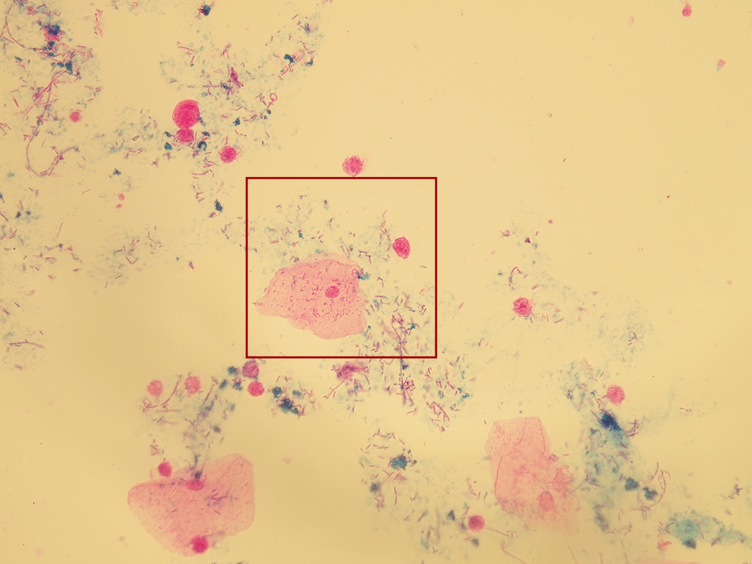
Bacteria inside squamous and transitional epithelia
Atypical cells
Occasionally, atypical cells may be found with urine sediment microscopy. In this case, the cells are probably of epithelial origin with variable shape, size and multiple nuclei. Such finding should be reported to the responsible physician. The patient was diagnosed with bladder cancer.

Atypical cells

Atypical cells