Erythrocytes (RBCs)
Erythrocytes are red blood cells without nucleus, with size about 6–7 µm and a disk-like shape. They count amongst the smallest and the most common elements in urine. Their presence in urine (hematuria) can be macroscopic or microscopic (without visible red color).
Causes for haematuria:
- Renal (glomerulonephritis, kidney cancer)
- Prerenal (hemocoagulation aberrations, muscle traumas, burns)
- Subrenal (bleeding in urinary tract – infection, kidney stones, carcinoma)
- Exertion (physical stress, cold)
If the erythrocytes have a normal biconcave shape with smooth surface, they are called eumorphic erythrocytes. Erythrocytes that passed to urine through glomerular membrane might be damaged and their shape is changed – we call them dysmorphic erythrocytes.
Dysmorphic erythrocytes may have a tire shape (codocytes) or the erythrocyte membrane may have protrusions (acanthocytes).
Hedgehog shaped or crenated erythrocytes (echinocytes) don’t count among the dysmorphic erythrocytes. They are deformed by erythrocyte dehydration in urine with high osmolality.
Stained sediment

Erythrocytes

Dysmorphic erythrocyte – codocyte

Dysmorphic erythrocytes – acanthocytes
Native sediment

Erythrocytes
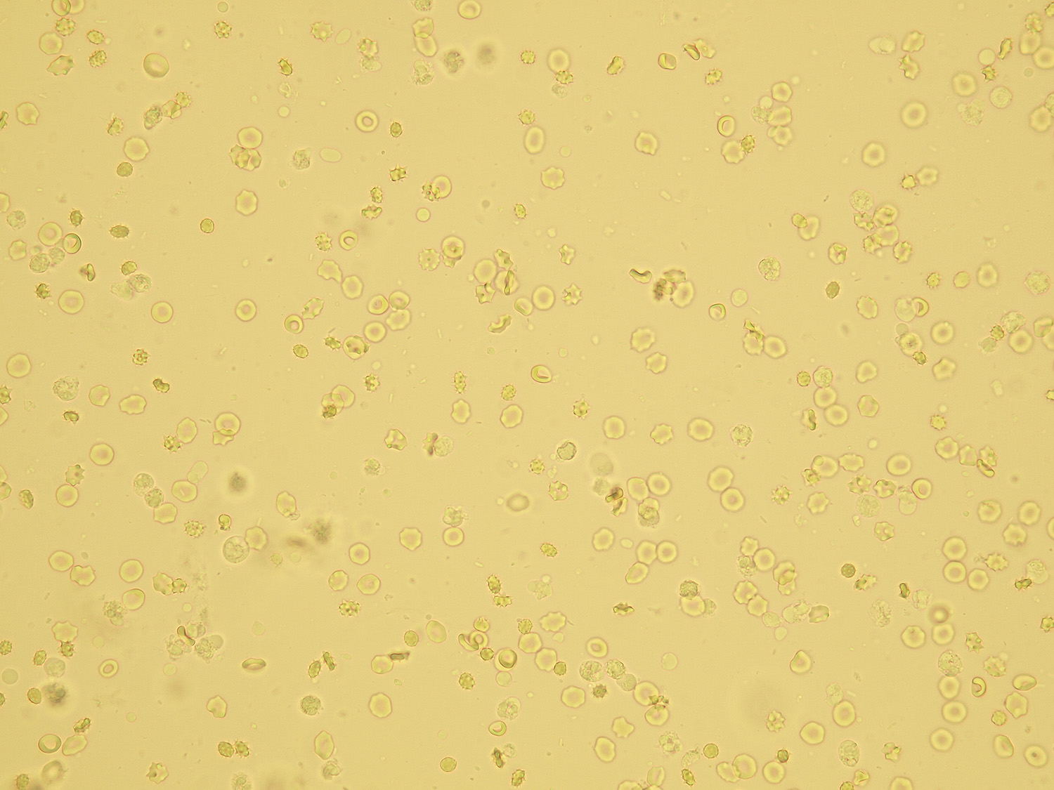
Erythrocytes deformed by high osmolality (the cells lose water)

Dysmorphic cells – codocytes

Dysmorphic erythrocytes – acanthocytes
Pictures from iQ 200 analyzer (IRIS)

Erythrocytes

Erythrocytes deformed by high osmolality (the cells lose water)

Dysmorphic erythrocytes