Strengthening of individual muscle groups
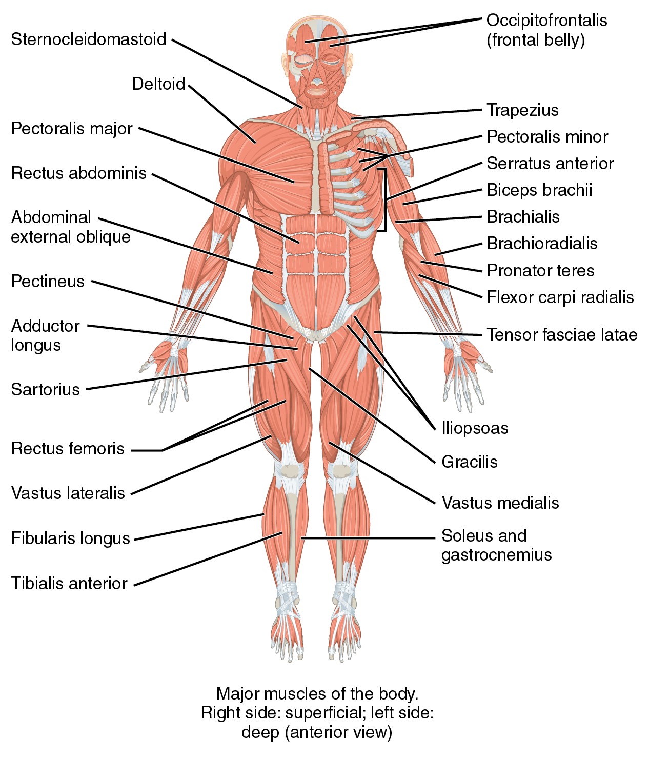
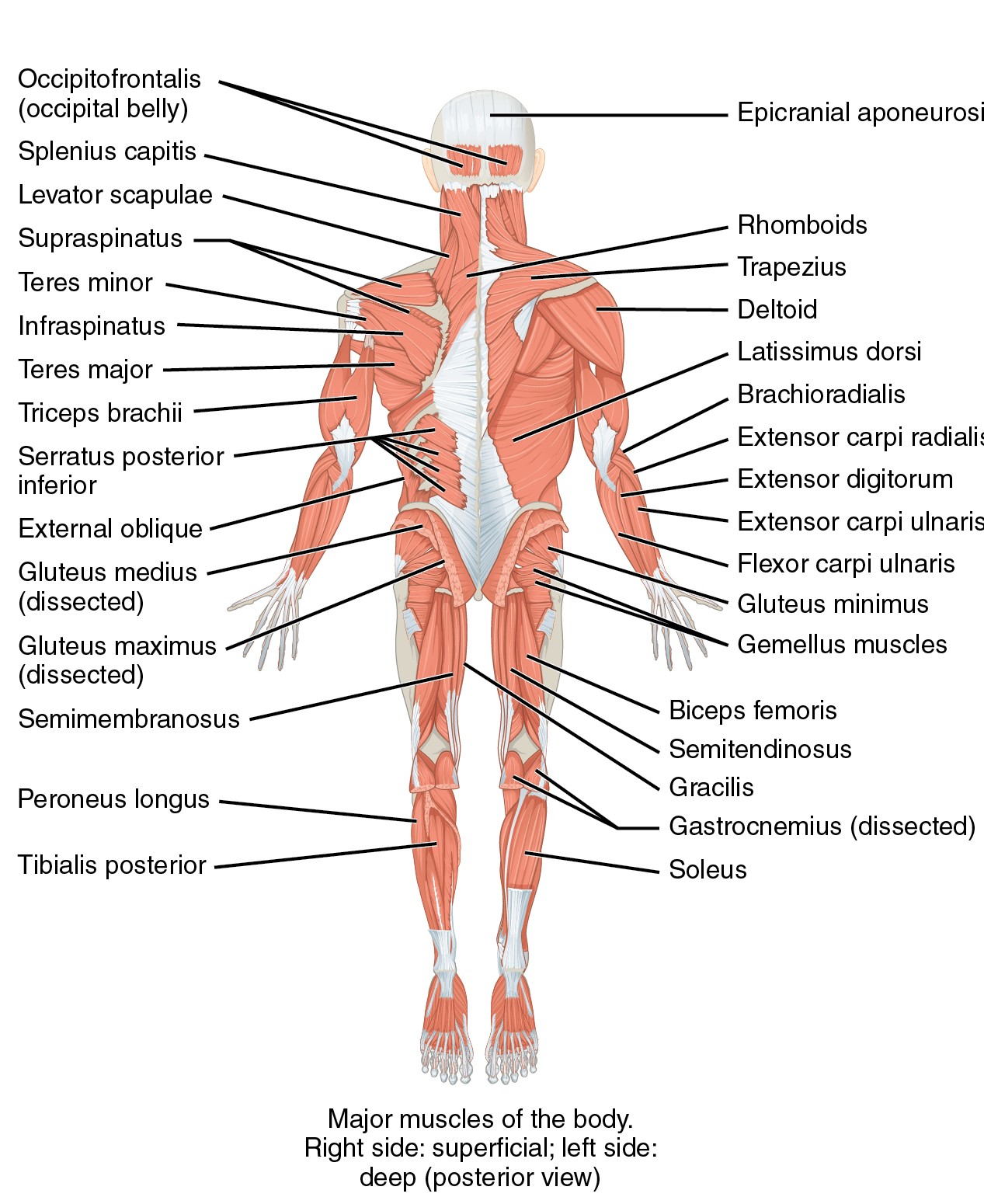
In this chapter there is a theoretical analysis of individual muscles and muscle groups, which are shown in Figures 17 and 18. We describe their function and how to strengthen these muscles. Following with Chapter 6, which describes the individual exercises used in bodystyling. For greater clarity, Chapter 6 is supplemented with photographs.
Strengthening the muscles of the legs and buttocks
The function of muscles of legs and buttocks
Leg muscles have a dual function - static and dynamic. They have almost the same configuration as the upper limb, but are stronger, bulkier, thicker and firmer. The big difference is in the femur and pelvic bone. The lower limb does not have as much freedom as the upper limb. The lower leg is missing the possibility of rotation, which is necessary in the forearm. The foot mainly has a static function so therefore doesn’t develop fine motor movements like the hand (Jarkovská, 2005).
Anatomy of leg muscles and buttocks
Muscles of the pelvic region
Iliopsoas muscle (flexion of the hip joint, muscle with a tendency to shorten)
- Hip flexion assists in pulling and external rotation of the lower limb.
- Rotates the pelvis and torso in the opposite direction.
- It is an assistant of gluteal muscles. Maintains the balance of the torso.
- We could not walk if the muscle would stop its function.
Gluteus maximus (muscle with a tendency to weaken) – is flat, the largest and strongest of the gluteal muscles. It has the largest formative effect on the buttocks. Walking low on a flat surface creates little activation. Its activity increases, for example, when climbing stairs, walking fast, walking in uneven terrain, running, and jogging. During the strengthening of the buttocks, the extension of the hip joint is most often used. Its healthy range is 10 - 15 °. If we exceed the range, then the lumbar flexors get involved (iliopsoas muscle and rectus femoris) and the lumbar spine extends excessively. If the trainee cannot tighten the core, then choose alternate exercises in supine position with the lifting of the pelvis. An even more effective exercise is at the kneeling position with the forearms down, by reaching the legs at the right extension without the pelvis moving. If we extent the leg at the knee, then we also strengthen the back of the thighs, if we bend the leg at the knee, we involve more of the gluteals. If a lot of repetition is included which make the muscle too tired, then the muscles of the lower back begin to engage. It can lead to pain of the hip and supportive leg. Due to the weakened medius gluteus, which does not hold the pelvis in the correct position. Together with gluteus muscles, hamstrings extend the hip.
Hamstrings and glutes perform hip extension.
- A fixed leg tilts the pelvis and thus ensures an upright position of the torso
- When bending forward it holds a predominant part of the weight
- Upper fibers abduct the thigh and lower fibers adduct it
Strengthening
The muscle tends to weaken, therefore it is necessary to strengthen it often in isolation, although it is very involved in all complex exercises. The upper part of the muscle is mostly involved in the maximum extension, the lower part is more influenced in exercises from a large initial flexion. Practicing involving gluteal muscles - lying on the back with knees bend, perform pelvic tilt (together with the abdominal muscles), this position cannot lead to inappropriate and very frequent conjunctions in the lumbar region.
Leg extension – there should be a simultaneous engagement of lumbar spinal extensors, which, however, is a highly fixed movement stereotype for many people – to eliminate the work of the lower back muscles the exercise is done in a supported kneeling position and the head hangs loosely, and the neck is flexed (reflexively also helps to stop using erectors). The heal leads the movement without simultaneous abduction (while standing, an external rotation can be used but the position is riskier due to the involvement of the lumbar region). The range of motion is determined by the possibility of extension in the hip joint –maximum of 15° (we can use gymballs to help the elimination of the work or lumbar region).
Squat – complex exercise significantly involving not only gluteus maximus muscle (mainly lower part), but also thigh muscles - flexors and extensors. Feet are shoulder-width apart or wider, for stability, feet are parallel, and knees are above toes. Exercise can burden the spine (mainly when using weights) – back is non rounded, erector spine must ensure the correct position of the spine. We should also pay attention to the knees – depth of the squat does not exceed 90° flexion in the knee joint.
Lunges forward and back – again very effective complex exercise, have the same principle as the squat, but the torso is held upright.
Gluteus medius – the outer side of the thigh (muscle with a tendency to weaken). It is located under gluteus maximus. It is a leg abductor and performs internal rotation of the hip.
Strengthening
Leg abduction – It is important to pay attention to the exact direction of movement, avoid external rotation of the thigh (toes up) or tilting pelvis forward followed by moving the leg forward (in this case the hip flexors take over the load). The bending of the standing leg should prevent the elevation of the pelvis, guiding the movement with the heel should avoid external rotation. Another option is lying on the side when the pelvis is fixed against elevation, the range of motion is greater than (45°).
Side lunges – significantly engage the muscles of the thighs (particularly the inner part), we pay attention to the upright posture with a neutral spine position, knee is always in line with toes.
Gluteus minimus – the outer thigh (with a tendency to weaken). It is hidden beneath the gluteus medius and is its helper. It is a leg abductor and performs internal rotation of the hip.
Strengthening – see gluteus medius
Tensor muscle of the fascia lata – the outer thigh (muscle with a tendency to weaken). Helps during the hip flexion, the leg abduction and influences the knee extension.
Thigh muscles
Sartorius muscle (muscle with a tendency to shorten) – this long, lean muscle originates at the hip bone and spirally goes diagonally down to the front of the thigh. Its insertion is on the inner side of the knee. Its functions are knee and hip flexion and external hip rotation.
Quadriceps femoris muscle – Its function is knee extension and hip flexion.
Strengthening
Except for rectus femoris muscle, this muscle tends to weaken. It is involved in all complex exercises.
Leg extensions – it is an isolated work in the knee, but only in the sitting position. Has 3 muscle heads with a tendency to weaken at the end of the extension (the last 10 to 15 °) therefore we are completing the move to full knee extension (the outer and inner head stabilizes the knee joint from the sides, therefore, the knee extension extends with small range of motion is included in the basic rehabilitation, e.g. after a knee joint operation) – also applies for squats.
Gracilis muscle – leg adductor – inner thigh (muscle with a tendency to shorten).
Leg adductor and knee flexor, lower leg rotates inward.
Adductor longus muscle and adductor magnus muscle – inner thigh (muscle with a tendency to shorten). Leg adductors and creates outer leg rotation.
Pectinate muscle –– inner thigh (muscle with a tendency to shorten). Leg adductor and hip flexor create outward thigh rotation.
Biceps femoris muscle – back and outer thigh, semitendinosus muscle and semimembranosus muscle – hamstrings – back and inner thigh (mucles with a tendency to shorten). All these muscles preform hip extension and knee flexion.
Strengthening
These are postural muscles and are shortened in most populations but at the same time they are weakened. They are sufficiently involved in complex exercises.
Leg curls – the effect can only be expected during resistance training
Lower leg muscles
Anterior tibial muscle, Extensor digitorum longus muscle, Extensor hallucis longus muscle – front side of the lower leg (muscles with a tendency to weaken) – all these muscles are extensors of the foot and toes. When they are weakened, it is difficult to pull the toes to the shin. They are engaged during walking, absorb shocks during jumps and can be strengthen by pulling the toes against resistance.
Peroneus longus muscle – outer side of lower leg (muscle with a tendency to weaken). Work during plantar flexion, secures the transverse and longitudinal arch of the foot.
Peroneus brevis muscle – outer side of lower leg (muscle with a tendency to weaken). Ensures internal rotation of the foot.
Triceps surae muscle (gastrocnemius and soleus) – back side of the lower leg (muscle with a tendency to shorten). The two heads have an origin at the lower end of the femur (gastrocnemius muscle) and the third head originates on tibia and fibula (soleus muscle –perform calf raises). They align with the Achilles tendon and the muscle insertion is on the calcaneus. The entire muscle is stretched down the leg and lifting the body during walking. The main function is to perform calf raises. The superficial part of the calf muscle flexes the knee joint.
Popliteus muscle, tibialis posterior muscle, flexor digitorum longus muscle and flexor hallucis longus muscle – back and inner side of the lower leg (muscles with a tendency to weaken). Performs plantar flexion and maintains the longitudinal arch of the foot.
Muscles of the foot
Located on the top and on the sole of the feet. Perform extension of the toes. The sole muscles bend and move the toes to the side.
Strengthening exercises
Complex exercises such as squats, semi-squat, lunges and similar movements act simultaneously on the gluteal muscles and the muscles of the thighs. By changing the position of the legs and torso the effect can be directed more on one or the other of the two muscle groups (E.g. a half squat or squat with the torso bending forward activate more the muscles of the buttocks).
Strengthening the abdominal and back muscles
Function of abdominal muscles
The problem area of the body is the abdominal muscles and muscles of the pelvic floor. Inactivity weakens the muscles quickly and ages them. In these areas, extra fat is easily stored. Abdomen bulges forward, lose their voltage and create a bubble. As a rule, this change leads to health consequence, back pain, mostly of the lumbar region. The solution is to start with these muscles to strengthen and stretch well.
Anatomy of abdominal muscles
Anterior and lateral abdominal wall consists of four pairs of muscles. The muscles are richly interwoven with flat tendons to strengthen the abdominal wall. They are stored between the chest and the pelvis. Every time the trunk moves, it engages all muscles at once, but in different proportions. They are working together as respiratory muscles. Abdominal muscles form a solid shell with a steady resting tension which keeps the organs in the abdominal cavity in the right position. Ensures proper bowel function, helps to empty the rectum, bladder and uterus. The abdominal muscles are a firm support for the lumbar spine. At any strain the muscles form a flat pillow on which the spine rests. Whenever we round the thoracic and lumbar spine, instantly the muscles shorten, the muscle contraction occurs, the chest is closer to the pelvis. The muscles are also maximally activated during rotation and side bends of the torso. Some parts of the abdominal muscles do not have the insertion to the bone but end in the ligaments and tendons of other muscles. Strengthening and stretching of the abdominal muscles is important for the correct posture (Jarkovská, 2005).
Rectus abdominis muscle is a long strap muscle that extends the entire length of the anterior abdominal wall. It is broader above and lies close to the midline, being separated from its fellow by the linea alba. It arises by two heads from the anterior aspect of the symphysis pubis and the pubic crest and it inserts into the 5th, 6th and 7th costal cartilages and the xiphoid process.
- Muscle flexes the vertebral column (torso).
- The obliques cooperate during flexion.
- When the pelvis is fixed, the lower part of abdominals is more active, the part attached to the pubic bone.
- When strengthening the abdominals lying on the back, it is always important to press the pelvis and the lumbar region to the ground.
Strengthening
The abdominal muscles are intertwined anatomically and functionally, during strengthening of the abdominal muscles we feel a sense of flattening the abdominal wall, drawing the ribs in the exhalation position and pressing the lumbar against the pad. No abdominal muscle passes the hip joint, that’s why the leg movement doesn’t activate abdominals but activates hip flexors. Strengthening abdominals should be included in all workout plans.
Chest/shoulder blades lift – engages the most upper part of the muscle that is practiced more for aesthetic than for health reasons. Basic crunches are done lying down with knees bent, feet on the floor. Hands are supporting the head without pulling the head up.
Pelvic tilt – the lightest form of exercise for the lower part of the muscle is the lifting of the underlying pelvis on the back with the support of the feet, lifting is performed only up to the lumbar region of the spine; in the case of enlarged thoracic kyphosis, head support is recommended.
Pelvic lift with legs up – execution is the same as the previous pelvic tilt, legs up adding to the resistance, legs should be stable (no kicking up or pulling legs to the head).
Obliques muscles
- Internal oblique muscle– origin: lumbar fascia, anterior two thirds of iliac crest and lateral two thirds of inguinal ligament; insertion: costal margin, aponeurosis of rectus sheath (anterior and posterior), conjoint tendon to pubic crest and pectineal line. Flexes trunk in a dual action, otherwise bends the torso to the side and rotates the torso.
- External oblique muscle – origin: anterior angles of the lower eight ribs; insertion: outer anterior half of iliac crest, inguinal ligament, public tubercle and crest, and aponeurosis of the anterior rectus sheath. Flexes trunk in a dual action, otherwise bends the torso to the side and rotates the torso.
Strengthening
Chest lift with rotation – starting position is the same as for crunches, we pay attention to the range of motion of the shoulder which moves diagonally to the opposite hip without lifting the pelvis.
Pendulum abs exercise – exercise for lower part of obliques (if the torso is fixed and the pelvis is rotating), leg up to the ceiling with knees bent (advanced level – straight legs), keep lower back on the floor, lower legs to one side and lift back up.
Side crunches – a motion containing a rotary and side bend component, the upper arm supports the head or is next to the torso, the shoulder approaches the hip, but the torso does not rise.
Standing side bend– apart from the oblique abdominal muscles, erector spinae muscles are activated; therefore we always compensate with enough stretching. The pelvis is not sufficiently stabilized in this position, the seated position would be better. The alternating side bends are less efficient since the loads are balanced. The side bend must be absolutely free of any slight bend forward, otherwise the function is taken over by the erectors.
Transverse Abdominal muscle – origin: costal margin, lumbar fascia, anterior two thirds of iliac crest and lateral half of inguinal ligament; insertion: aponeurosis of the posterior and anterior rectus sheath and conjoint tendon to pubic crest and pectineal line; function: supports abdominal wall, aids forced expiration and raising intra-abdominal pressure. Conjoint tendon supports posterior wall of inguinal canal. Its function is to help with correct posture and correct technique during abdominal exercises.
Strengthening
Isometric strengthening of abdominal muscles – our approach to strengthening the abdominal muscles is based on their dynamic function, but in everyday life we almost never use this function. Far more significant is the static function of the abdominal muscles = maintain abdominal pressure and active engagement in maintaining proper posture, the principle applies that the muscle adapts to the function it performs, that is, we cannot expect dynamic exercise to greatly improve static functions. Static function training can be performed in several positions but suitable for beginners are those in which the muscles do not have to act as antigravity., for example:
1. lying on the stomach we try as much as possible to draw the abdominal wall inward while maintaining a neutral spine.
2. in supine position, knees bent we pull the stomach in without the pelvis tilting (we need to practice the involvement of abdominal muscles in a neutral position).
3. planks, hovers and other balance, unstable positions are challenging, so it is important to always pay attention to the neutral spine posture and regular breathing – the goal of the exercises is by repeated conscious training convert this activation of the abdominal muscles to the subconscious and apply it in everyday life, thus improving posture and spinal relief.
Oblique and transverse abdominal muscles play an important role in breathing. During exhale they pull the chest down, increasing the intra-abdominal pressure and pushing the diaphragm upwards. In case of inhalation, they fixate the lower ribs and help to lower the diaphragm. The abdominal muscles ensure that each segment of the spine is bent against one another. The function of the abdominal muscles is the kyphosis (rounding) of the spine.
Strengthening of abdominal muscles
When strengthening, a position is selected where we do not activate hip flexors and the muscle in the lumbar region (lying with knees bend…). During exercise, we try to pull the abdominal wall in, the movement is carried out with each exhale, we pay attention to proper head fixation – the head is in a slight inclination, chin bent towards the chest, the crown pulled out into the distance in the extension of the chest, we pull the shoulders away from the ears.
Function of back muscles
On the back of the torso there is a group of muscles, which is spread in four layers. The muscles of the limb origin are placed on the surface, first and second layers. They begin on the spine and are clamped on the shoulder blade or humerus. In the third layer, the muscles are spread from the spine to the ribs. The deep fourth layer is composed of complex back muscles.
Determining the exact function of the back muscles is difficult and often inaccurate. In practice, we most often use the term for muscles that we strengthen the name of the lower blade fixators. These are the muscles that connect the shoulder blade with the spine or ribs. They work during movements of the shoulder joint. If these muscles are strengthened, then our backs are straight, and the shoulder blades are not protruding. In conjunction with the upper blade fixators, they fix the shoulder blade and prevent excessive movement of the upper limb in the shoulder joint. Among the lower fixators include rhombic muscles and the lower and middle part of the trapezius muscle.
The first (surface) layer:
Trapezius muscle – has a descending (upper) and ascending (lower) portion. It pulls and fixes the shoulder blade to the spine and pulls the shoulders back. The upper part lifts the shoulder blade, the lower part pulls the shoulder blade down. Both trapezius muscles tilt the head back, and the contraction of one muscle tilts the head to its side.
Strengthening
Strengthening refers to the middle and lower part of the trapezius muscle, it is necessary to explain some terms used for the muscles around the shoulder blades.
Rhomboideus muscles = the middle part of trapezius (horizontal fibers), minor and major rhomboideus muscle
Lower shoulder blades fixators = lower part of trapezius (ascending fibers), part of latissimus dorsi (horizontal fibers) and serratus
External arms rotators = infraspinatus and teres minor – interplay of these three muscle groups keeps the shoulder blade in the correct position – they attract to the spine (adduction), they press it against the chest and compress it downwards (depression).
Shoulder blade adduction with external rotation
Also involved in a number of exercises for latissimus dorsi and back of the deltoid muscle
Latissimus dorsi – action: arm adduction, moving arm behind, pass the torso and rotates humerus in the shoulder joint inward. Pulls shoulder down. It is an auxiliary breathing muscle.
Strengthening
Lat pull down – an error is the guiding motion with bending elbows and head pretension, we concentrate on pulling of the shoulder blades down in the final phase, option to pull the bar to the chest allows more simultaneous activation of rhomboids and lower blades fixators.
Bend over row – always pay attention on keeping the spine straight and the head in its extension. Exercise can be done both with two-handed and one-handed with support which is easier on the spine and includes the upper trapezius, also involve rhomboids and lower shoulder blade fixators (particularly the variant with the elbow away from the body – there is also the risk of involvement of the upper trapezius, therefore we monitor the lifting of the shoulders) and external arm rotators (especially in a variant with external rotation, that is, turns thumbs away from the body)
Double arm bend over row - exercise for advanced exercisers because of static engagement of erector spinae, which must be sufficiently strong enough. The variant with overhand grip is that all back muscles are involved, during the variant with underhand grip hands are closer together, the movement is toward the waist and stimulates primarily the lower and inner part of the latissimus dorsi together with other back muscles. During the variant with underhand, narrow the grip and elbows away from the body, the upper latissimus dorsi and rhomboids are more involved.
Rows bottom – “rowing” It is done properly by pulling the shoulder blades to the spine and by ejecting the sternum, it involves in particular the outer part of latissimus dorsi and the lower mid-scapular fixators. In the initial position of bending forward it activates the spinal extensors, especially in the lumbar region.
Second layer:
Rhomboids major and minor (lower shoulder blade fixator) – pulls shoulder blade towards the spine.
Strengthening – see muscles between shoulders
Levator scapulae (upper shoulder blade fixator) – lifts shoulder blade and pulls it toward the spine. When shoulder blade is fixed, it bows the cervical spine to its side and slightly backward.
Strengthening
It is part of the postural muscles, that’s why we mainly stretch it. Along with the upper part of the trapezius it works as an upper shoulder blade fixator. This function is excessively active in everyday life and is often inconsistent with the function of the lower shoulder blade fixators.
Third layer:
Serratus muscles (lower shoulder blade fixators) – raises the upper ribs, fixes and pulls the lower ribs. It is an auxiliary breathing muscle.
Fourth (deep) layer:
It consists of a complicated complex of back muscles. Muscles extend longitudinally from the sacrum to the occipital bone. It performs the fixation function – creates a muscular corset around the spine, and dynamic function – ensures the movement of vertebrae, rotation, side, forwards and backwards bend. The muscles placed deeper and closer to the spine are shorter. The deepest muscles connect adjacent to the vertebrae. All these muscles straightening the torso, are named as head and spinal extensors. During unilateral contraction the spine bends sideway, during bilateral contraction they straighten the torso, pull the ribs down and prevent them from lifting. To strengthen them, we usually choose symmetrical exercises, in which the torso is elongated or rotated around the longitudinal axis. To gain strength, these exercises are effective in the unstable positions, for example, strengthening on a big ball.
Strengthening
Although it is a postural muscle, we should devote ourselves to sufficient stretching of these muscles, they also must be strong enough, which, especially in the lumbar region, is not always the case.
Torso extensions – performed only to the position where the torso is in line with the thighs, not to hyperextend, which fixes the wrong position of the lumbar spine.
Strengthening of the back muscles
If the starting position is lying on the stomach, it is recommended to slightly lift the hips off the ground and tuck the abdomen. This will prevent excessive curvature in the lumbar spine and that will create greater involvement of the thoracic spinal extensors.
Strengthening muscles of the neck, chest and arms muscles
Function of the neck muscles
The muscles of the neck lie between the skull, the spine, and the chest. They tend to weaken, that is why we have to do both, stretch and strengthen them. In order for the neck to fulfill its supporting role, it is necessary to create a cervical corset from the muscles around the cervical spine. Neck muscles engage in both simple and complex movements, e.g. during exercises for abdominal and back muscles while lying on the back and on the stomach. For all the exercises, where we emphasize "straight back, do not lift your shoulders", we strengthen the muscles of the neck. All forward bends, as well as head and neck rotation are practiced in a slow and smooth pace by alternating between tension and relaxation (Jarkovská, 2005).
Anatomy of the neck muscles
Sternocleidomastoid muscle – Function: to flex, lateralize and rotate the neck.
Scalene muscle – Function: helps to fix the position of the neck and to move it laterally.
Strengthening
Neck muscles have a tendency to shorten but that doesn’t mean that they should only be stretched – firm muscles create a protective corset around the cervical spine. As a special exercise, to make them strong, we should do isometric exercises against a solid support from the side, and front and back (e.g. car headrest). Exercises are carried out with care and the correct posture of the head is observed.
Head flexion – stretching the muscles on the back of the neck.
Function of chest muscles
In the shoulder joint, the major actors involved are the deltoid muscle, the large pectoralis muscles, and the trapezius muscle. The main function of the shoulder girdle muscle is the movement in the shoulder. Breast muscles look aesthetically if they are well developed. The arm muscles are attached to the shoulder girdle or to the humerus, the pectoralis are attached to the sternum. The diaphragm has a special position.
The first layer:
Pectoralis major – are primarily responsible for the movement of the shoulder joint. The first action is flexion of the humerus, secondly, it adducts the humerus. When contracted it lowers the arm when it is raised and, if it is already lowered, then it moves the shoulder forwards, and bending it back.
Strengthening
Upper pectoralis originating on the clavicle is usually strong enough (used during arm raise forward, which is a frequent movement in everyday life – while typing on the keyboard, moving the arms while walking, etc.), the middle part of pectoralis major (horizontal fibers starting at the sternum) is weakened, activated during horizontal adduction, the lower part of pectoralis is usually shortened.
Chest fly – this exercise activates the horizontal fibers of the pectoralis. Movement starts with arms in front of the chest with elbows slightly bent, bend elbows more during arms opening to the side, palms face each other, the movement ends when the palms are at shoulder level.
Chest press – the exercise again acts on the middle part of the pectoralis muscle. Both initial and final positions are the same as for chest fly, except the palms are facing forward this time. It can be practiced with one-handed dumbbells or with a bar.
Modified peck-deck – elbows position relative to the shoulders position affects the involvement of the individual muscle parts therefore, the base position of the elbow is at shoulder height. The movement starts with the elbows at the level with the shoulder axis and ends with the elbows approaching the center of the body. The exercise efficiency can be increased by internal arm rotation (little finger edge forward).
Pectoralis minor – is located under the pectoralis major, pulls the shoulder blade forward and down, helps to lift the ribs. It also acts as an auxiliary respiratory muscle.
Serratus anterior muscle – moves the internal border of the scapula forward and elevates the shoulder. In addition, it has respiratory functions, elevating the ribs and widening the thorax.
Strengthening – see muscles between shoulder blades and deltoid muscle
Second (deep) layer (own chest muscles)
Intercostal muscles internal and external with the function of respiratory muscles
Diaphragm – flat muscle which separates the thoracic cavity from the abdominal cavity.
Function of upper limb muscles
We divide the upper limb muscles into shoulder muscles and shoulder blade muscles, arm, forearm and hands muscles. By neglecting to exercise the arms, their mobility and strength will be reduced. Muscles and ligaments are shortened, stiffened and become weak. The muscles in the shoulder girdle connect the arm with the shoulder blade and the collarbone. Fast atrophy is most common in people with a sedentary lifestyle. Any movement that their weakened arm muscles do is inadequate and has a low range of motion. This muscle weakness manifests itself in that they do not have the power to move with their own body repeatedly.
Muscles of shoulder and shoulder blade
Deltoid – it consists of three parts. The front, middle and back heads. It is the most massive flat muscle that wraps and models the shoulder joint from all sides. It keeps its resting tension on the humeral head and keeps it in the hole in the shoulder joint. The anterior and posterior part of the muscles allows for the front or back raise, the middle part lifts the arms to the side.
Strengthening
While the front part of the muscle is usually hypertrophied, the middle and rear part are weakened.
- The front part:
Front raise with dumbbells – the position of thumbs facing each other activates the desired engagement of the middle part of the muscle (position of palms facing each other activates only the front part of the muscle)
Overhead press with bar – the bar is lowered only to the level of the last cervical vertebra, the grip is slightly wider than the shoulder width (wide grip is not suitable for overloading the shoulder joint), keep the head in line with the spine – avoid moving the head forward, and the exercise is not recommended for beginners.
Pull ups – unsuitable for beginners because of the activation of the upper trapezius, the grip width is about 20 cm, when the palms are drawn to the neck and the elbows are pulled back in the upper part of the movement then the posterior part of the deltoid muscle is involved, in addition to the anterior and middle parts.
- The middle part
Arms abduction – the movement ends when the palms are at shoulder level or slightly above them, moving the arms further up usually leads to the activation of the upper trapezius, the variant with internal arm rotation engages more rear and middle part of the deltoid, the most common mistake is lifting the shoulders and bending the torso towards the back.
- The back part
Is involved in the external rotation and arm extension, yet it is recommended to strengthen it in isolation – it contributes significantly to the posture in the area of the shoulder blade and shoulder joint.
Arm abduction while leaning forward – it can be done standing and sitting, movement leads through the back of the arms, a suitable option is to exercise only one arm (second arm supports to prevent torso rotation). For better concentration and partial relief of lumbar back, lying down on the side also activates the muscles between the shoulder blades and completely eliminates the burden on the spinal extensors.
Below the posterior part of the deltoid muscle are supraspinatus and infraspinatus muscles. The muscles help in abduction and external rotation of the arm.
Teres minor and major and subscapularis muscles help in arm adduction and perform external and internal arm rotation.
Strengthening – see the mid-scapular muscles and trapezius
Arm muscles
Biceps brachii – bends the elbow joint and externally rotates the forearm (supination), helps to lift the arms to the front, side and back.
Strengthening
Triceps extensions – the upper arm remains in the same position, extension is done with the elbow only, keeping elbows close to the head –the long head is involved more; when the elbows are open more – the other two heads are involved. Using a fast movement is a music, especially during releasing when the elbow joint is overloaded.
Triceps press – triceps bench press with underhand grip – works on triceps and pectoralis muscles
Kick-back – provides support to the torso with the hand on the thigh. The working arm is extended back with elbow bend, elbow is kept close to the body, only the forearm is moving.
Triceps dips – the body must be in a vertical position (the back is just behind the edge of the raised pad), range of motion is given by the joint.
Forearm muscles are divided into 3 groups:
The anterior group of muscles flexes the elbow joint, wrist and fingers, and rotates the forearm in.
Lateral group performs wrist extension and rotates the forearm outside.
Back group extends wrists and fingers.
Muscles of the hand
Muscles of the hand complement the functions of the forearm muscles, mobility of the thumb and little finger, expand and attract fingers.