Faculty of Medicine, Masaryk University
RNDr. Miroslava Beňovská, Ph.D., Mgr. Ondřej Wiewiorka, MUDr. Jana Tůmová
Crystals
The presence of crystals and amorphous microcrystalline deposits in urine is not considered a significant clinical finding. However, both parameters are determined and their amount is evaluated.
Crystals have various crystalline structure and they occur in many forms. The pH of urine is important factor for their formation and structure, although it is sometimes difficult to distinguish them even then. In that case, the elements could be classified only as crystals – without further specification.
The most frequent crystals in urine are: oxalate and uric acid (in acidic urine) or phosphate (in alkaline urine). Rarely found crystals in urine: bilirubin, cysteine, leucine, tyrosine or drug. We distinguish two types of amorphous microcrystals – amorphous urates in acidic urine and amorphous phosphates in alkaline urine.
Oxalates
Stained sediment
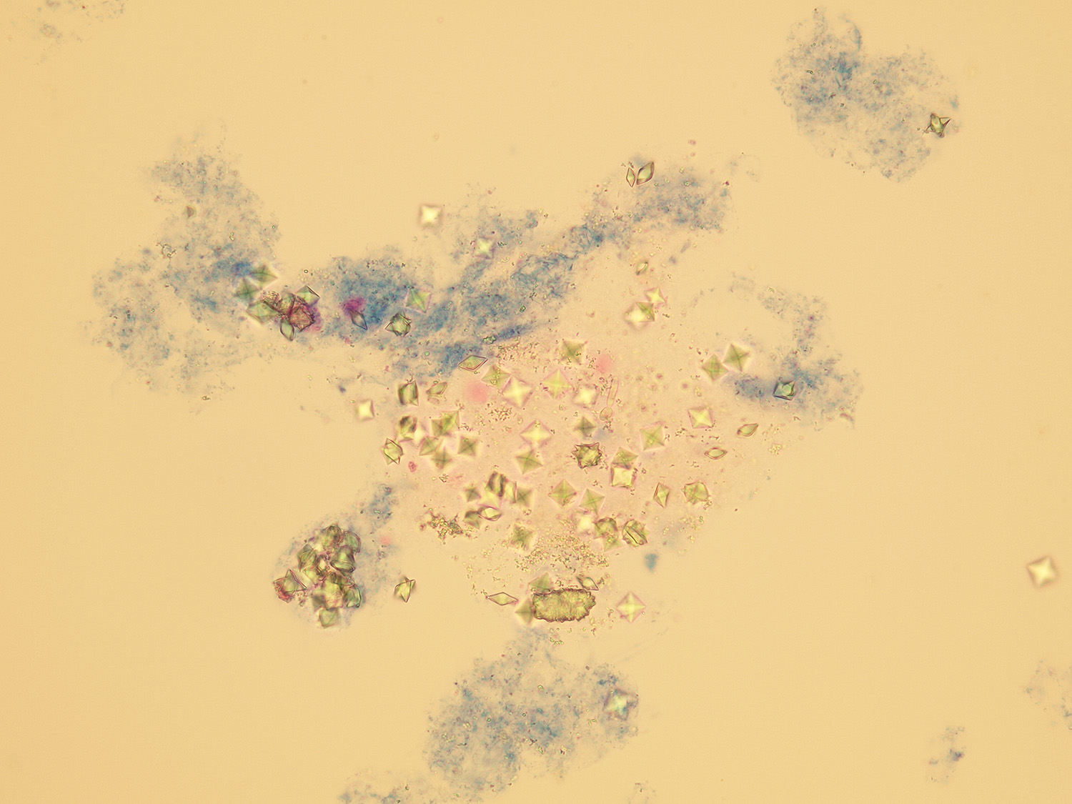
Calcium oxalate dihydrate (envelope-shaped)

Calcium oxalate monohydrate (oval and biscuit-shaped)
Native sediment

Calcium oxalate – monohydrate (oval form) and dihydrate
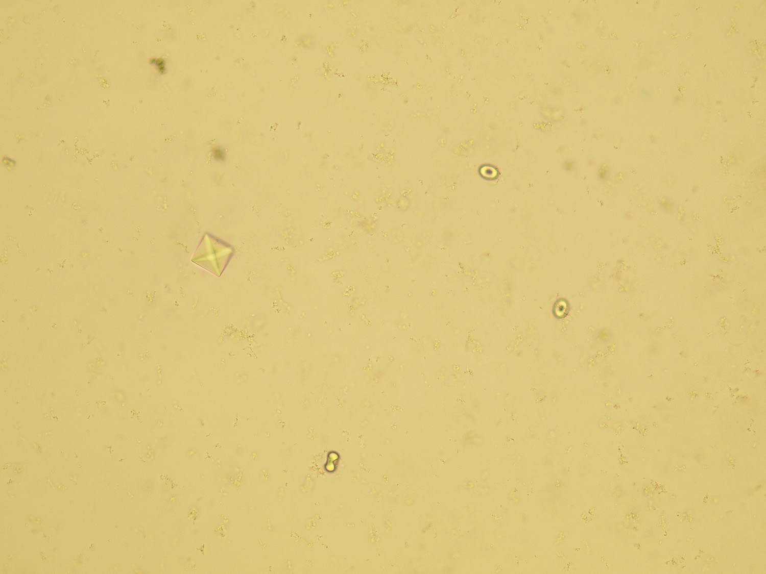
Calcium oxalate – monohydrate (oval form) and dihydrate
Pictures from iQ 200 analyzer (IRIS)
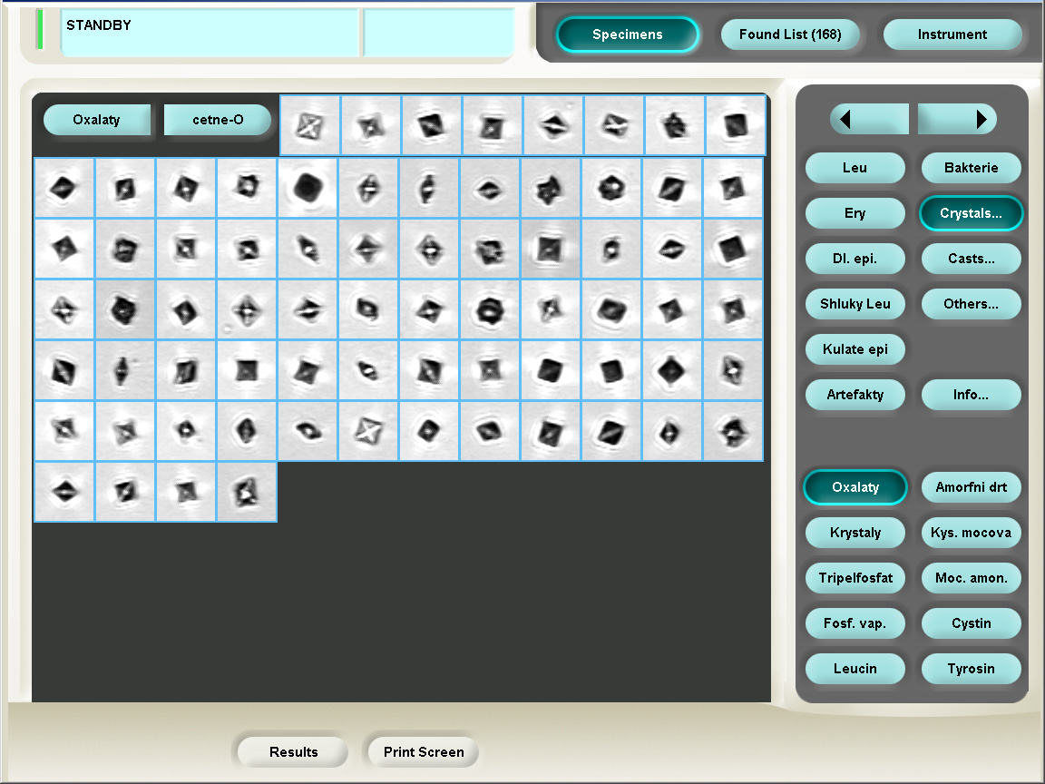
Calcium oxalate dihydrate (envelope-shaped)

Calcium oxalate – monohydrate (oval form) and dihydrate
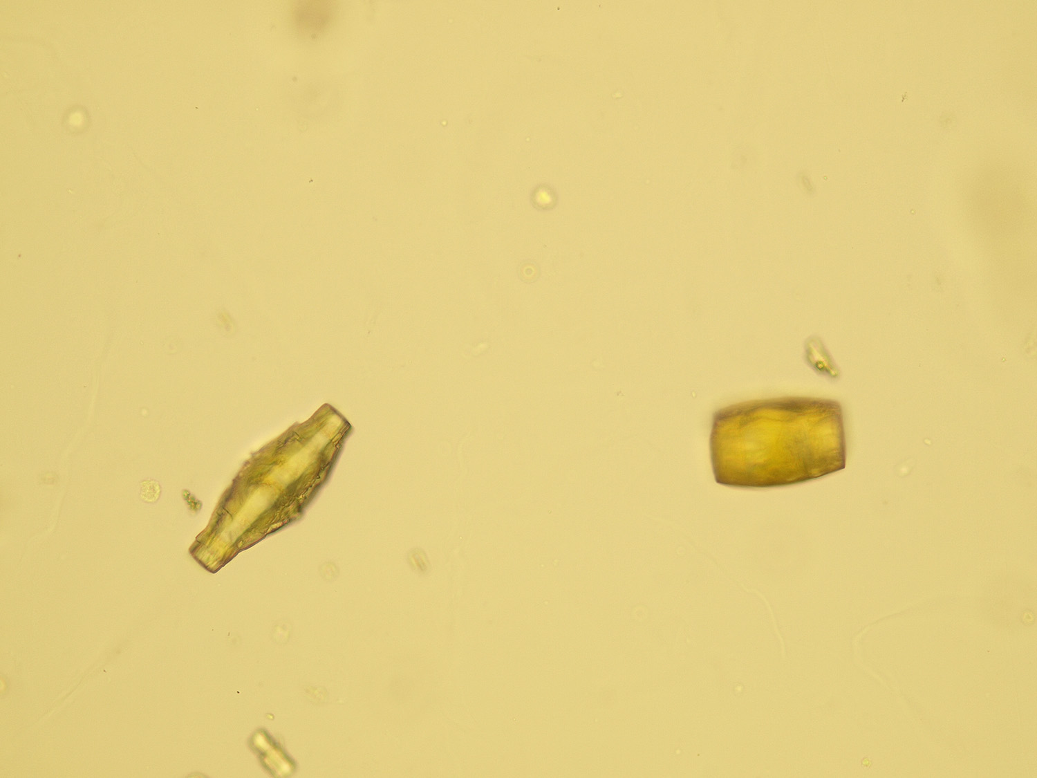
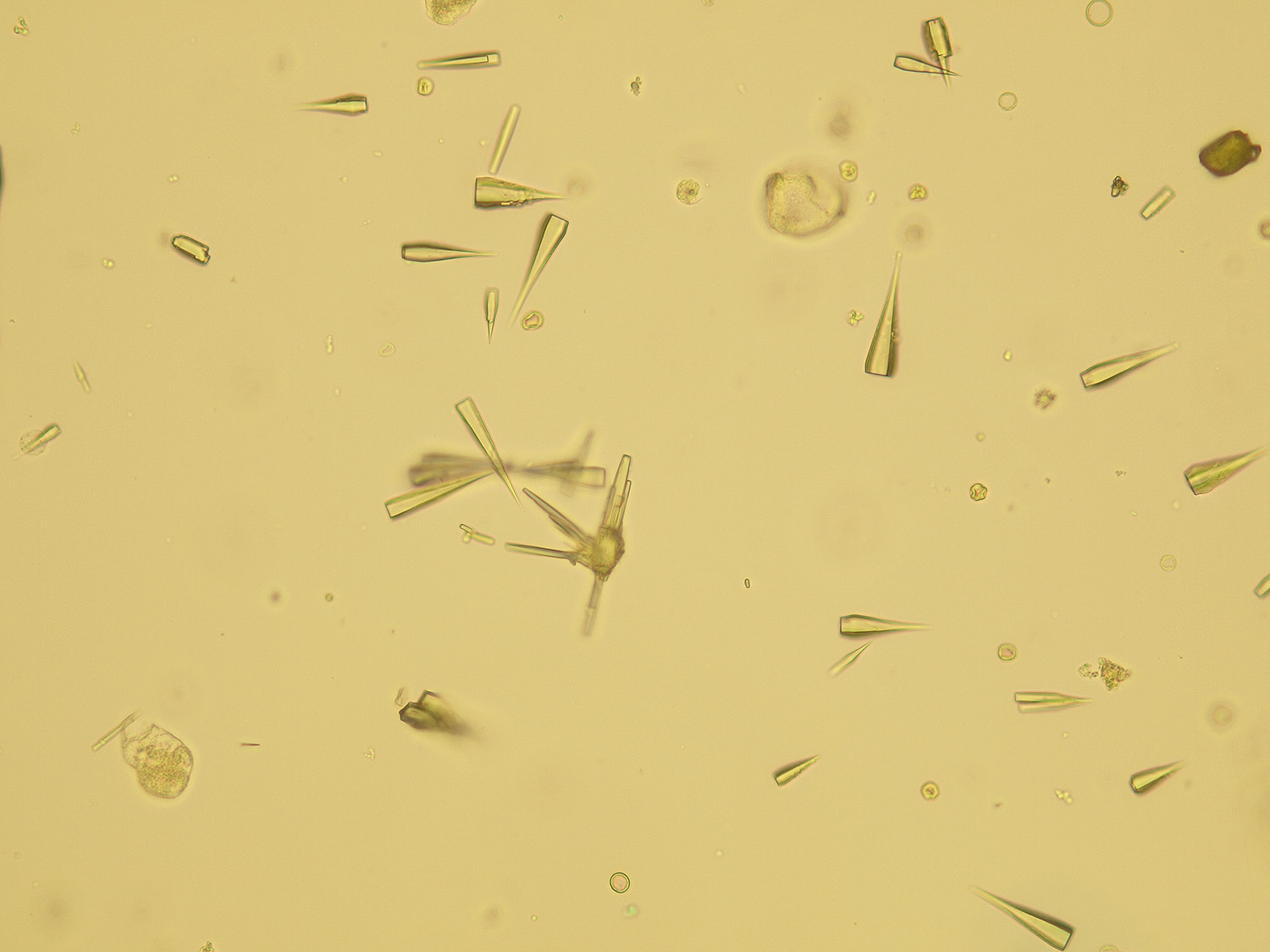
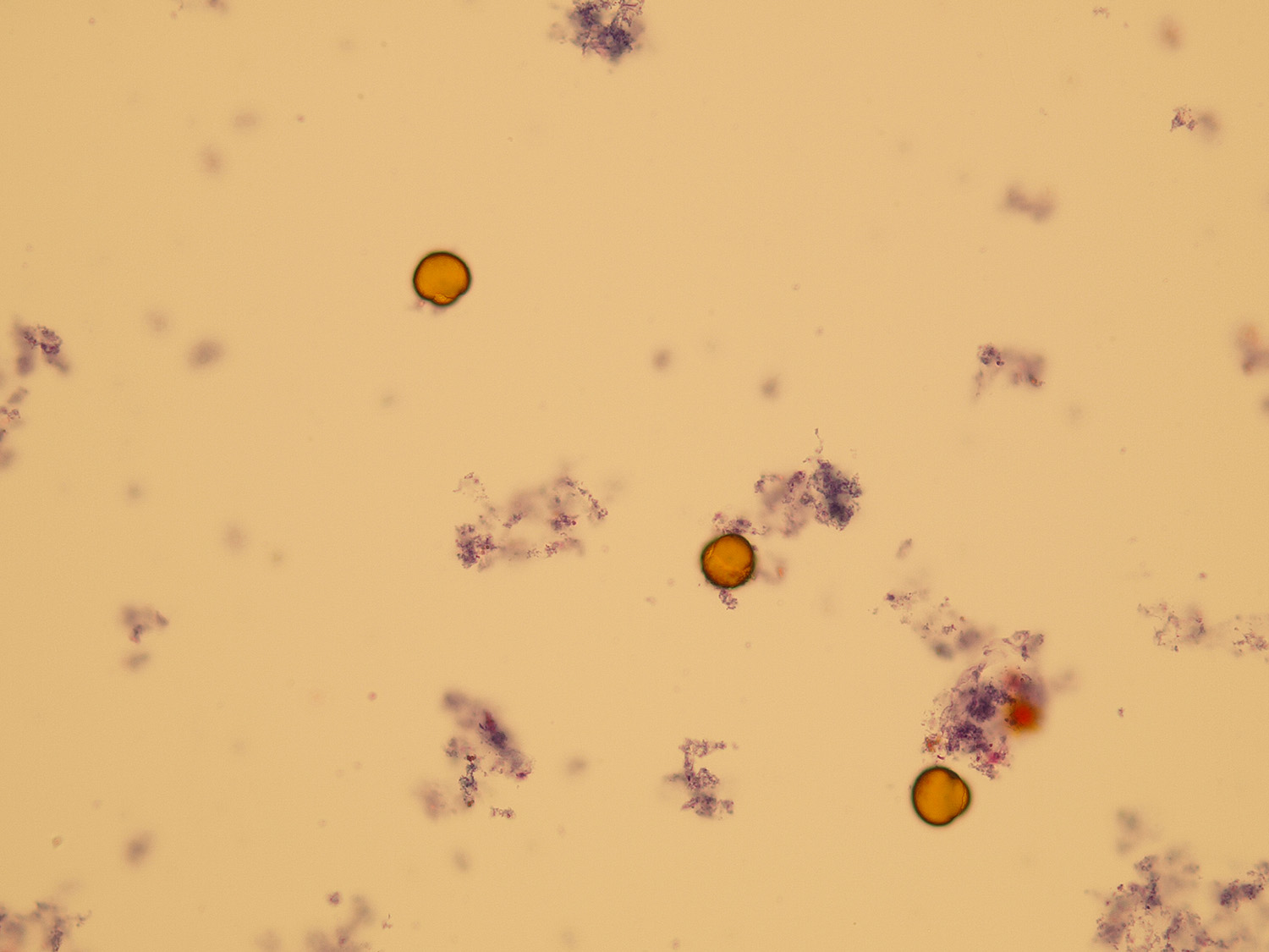
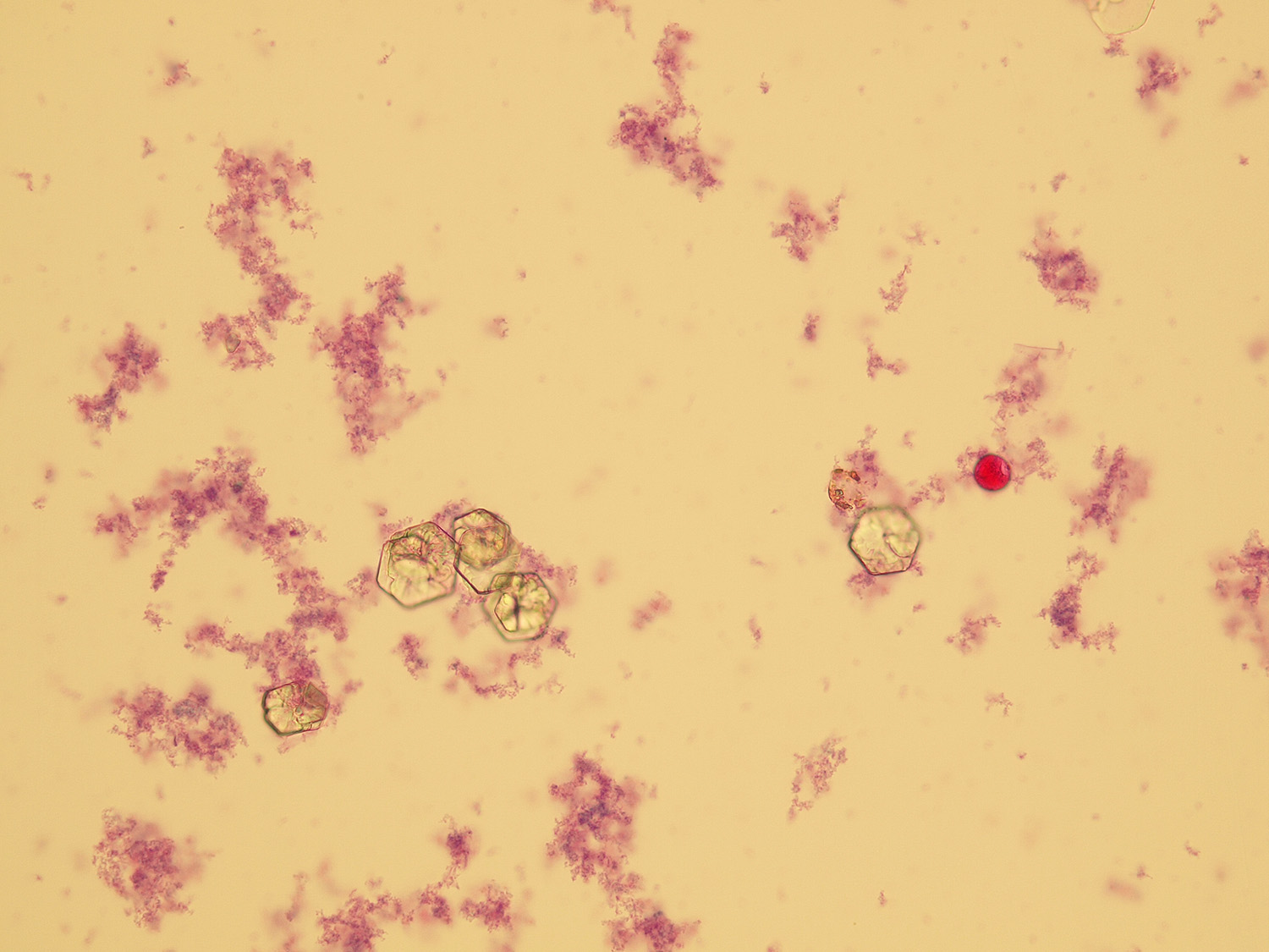
Uric acid
Various forms of crystals of uric acid
Stained sediment
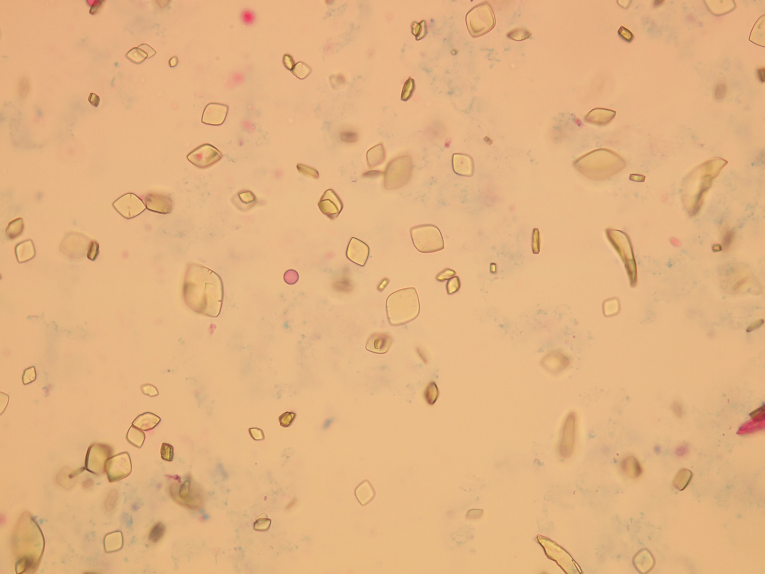
Uric acid (lemon-shaped)

Uric acid (barrel-shaped)

Uric acid (needle form)

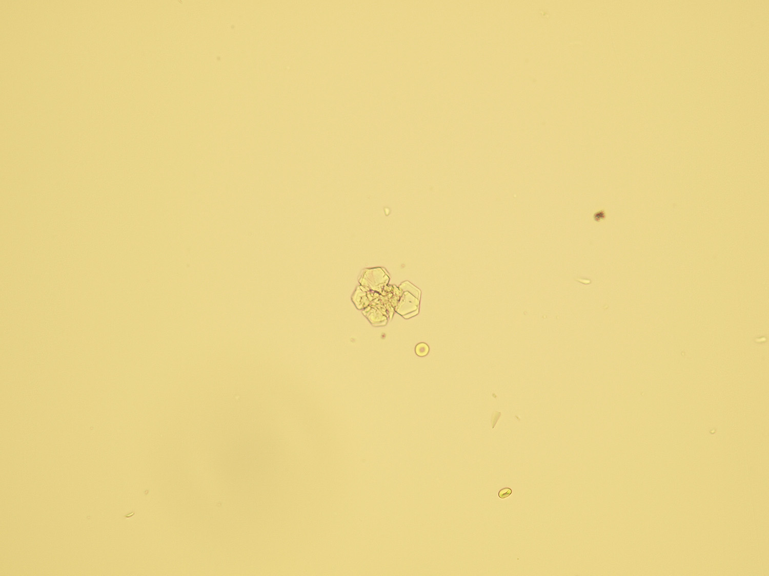
Uric acid
Native sediment

Uric acid (lemon-shaped)
Uric acid (barrel-shaped)
Uric acid (needle form)

Uric acid
Pictures from iQ 200 analyzer (IRIS)

Uric acid (lemon-shaped)

Uric acid

Uric acid (needle form)

Uric acid
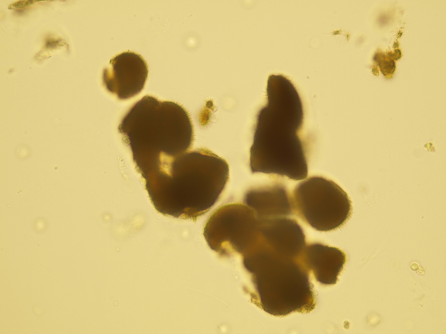
Ammonium urate
Stained sediment
Ammonium urate

Ammonium urate
Native sediment
Ammonium urate
Pictures from iQ 200 analyzer (IRIS)

Ammonium urate

Ammonium urate
Triple phosphate
Ammonium magnesium phosphate crystals (Triple phosphate).
Stained sediment

Triple phosphate (coffin-shaped)
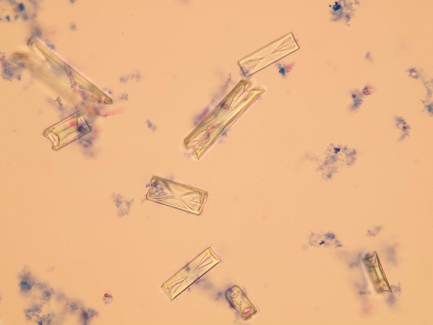
Triple phosphate

Triple phosphate crystal

Triple phosphate (arrows)
Native sediment
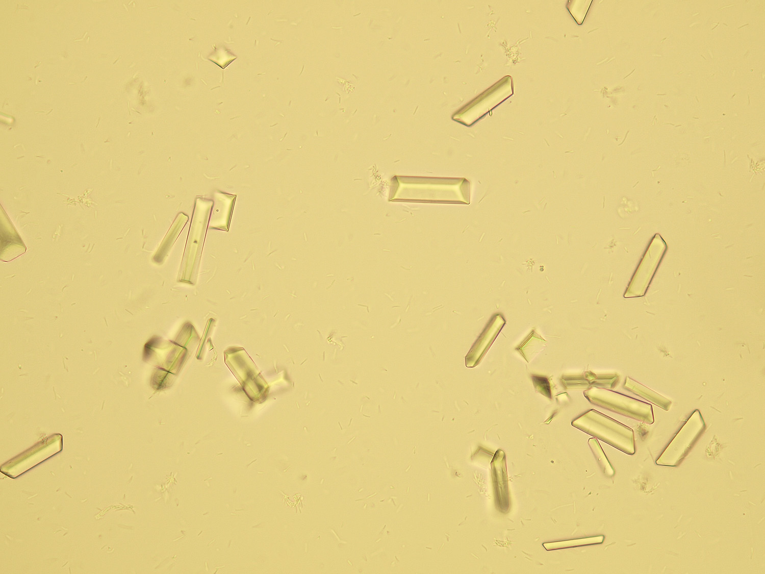
Triple phosphate (coffin-shaped)

Triple phosphate

Triple phosphate

Triple phosphate (arrows)
Pictures from iQ 200 analyzer (IRIS)

Triple phosphate (coffin-shaped)
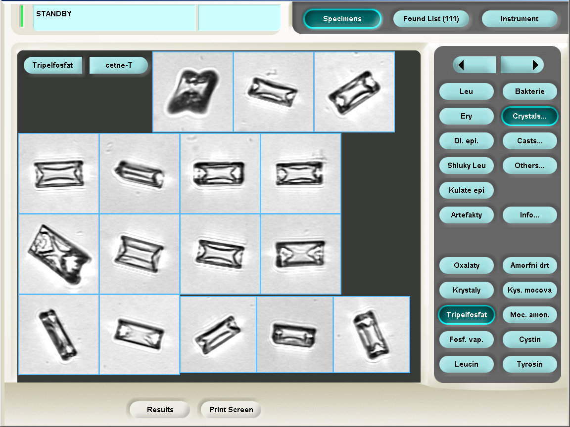
Triple phosphate

Triple phosphate

Triple phosphate

Massive triple phosphate
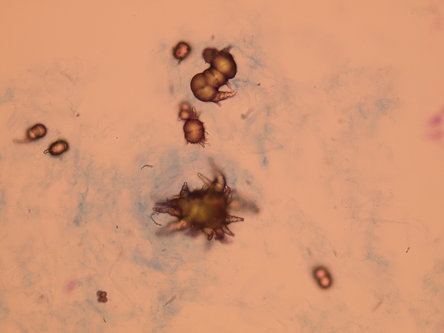
Calcium phosphate
These crystals can be differentiated from uric acid crystals by polarization microscopy. Unlike uric acid, calcium phosphate doesn't turn polarized light.
Stained sediment

Calcium phosphate

Calcium phosphate (needle-shaped and star-shaped druses)

Calcium phosphate
Native sediment

Calcium phosphate

Calcium phosphate (needle-shaped and star-shaped druses)
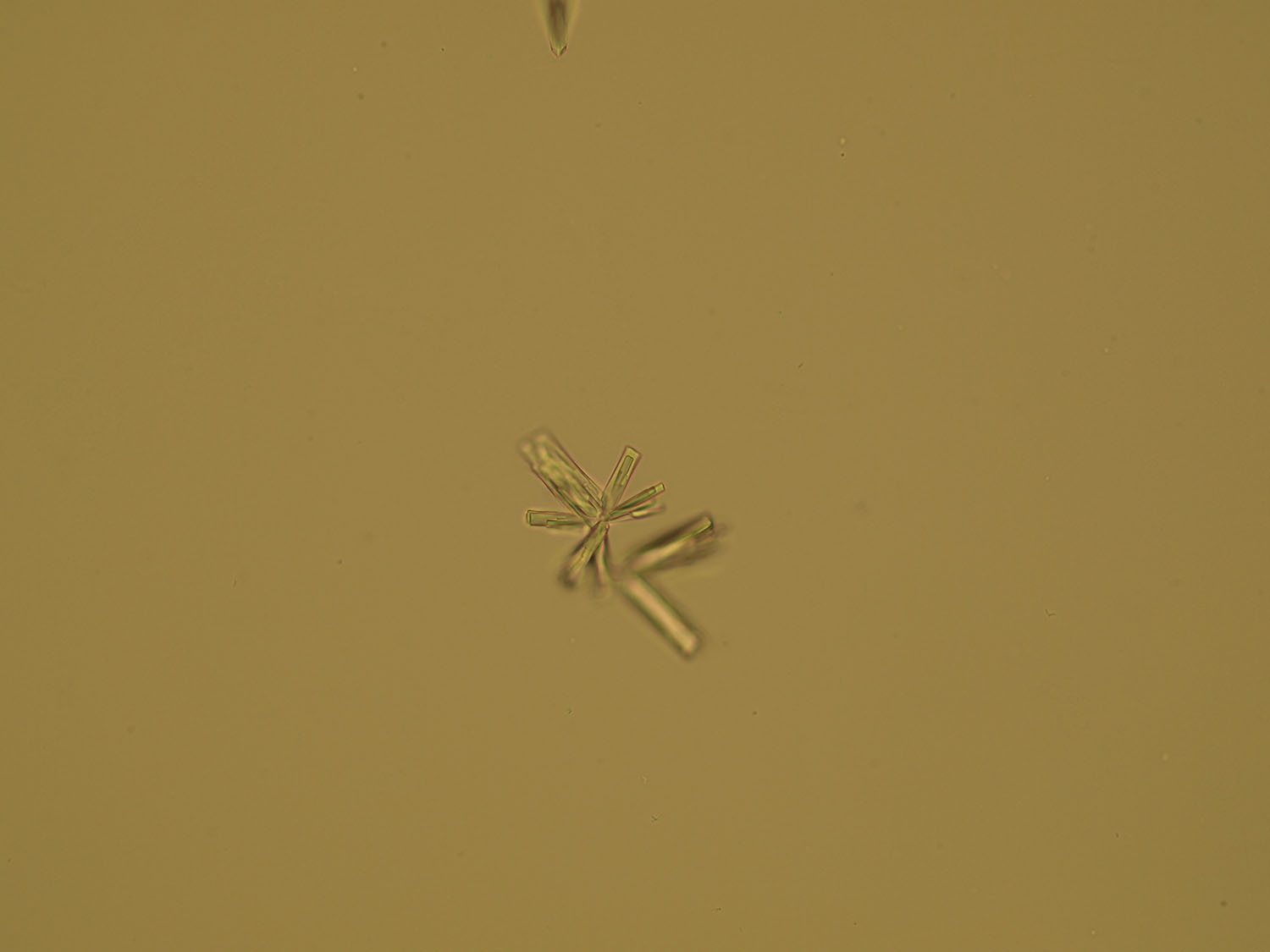
Calcium phosphate
Pictures from iQ 200 analyzer (IRIS)

Calcium phosphate

Calcium phosphate (needle-shaped and star-shaped druses)
Bilirubin
Stained sediment

Bilirubin
Bilirubin
Native sediment

Bilirubin

Bilirubin
Pictures from iQ 200 analyzer (IRIS)

Bilirubin

Bilirubin
Cystine
Stained sediment

Cystine
Cystine
Native sediment

Cystine
Cystine
Pictures from iQ 200 analyzer (IRIS)

Cystine

Cystine
Amorphous microcrystals
Stained sediment

Amorphous microcrystals
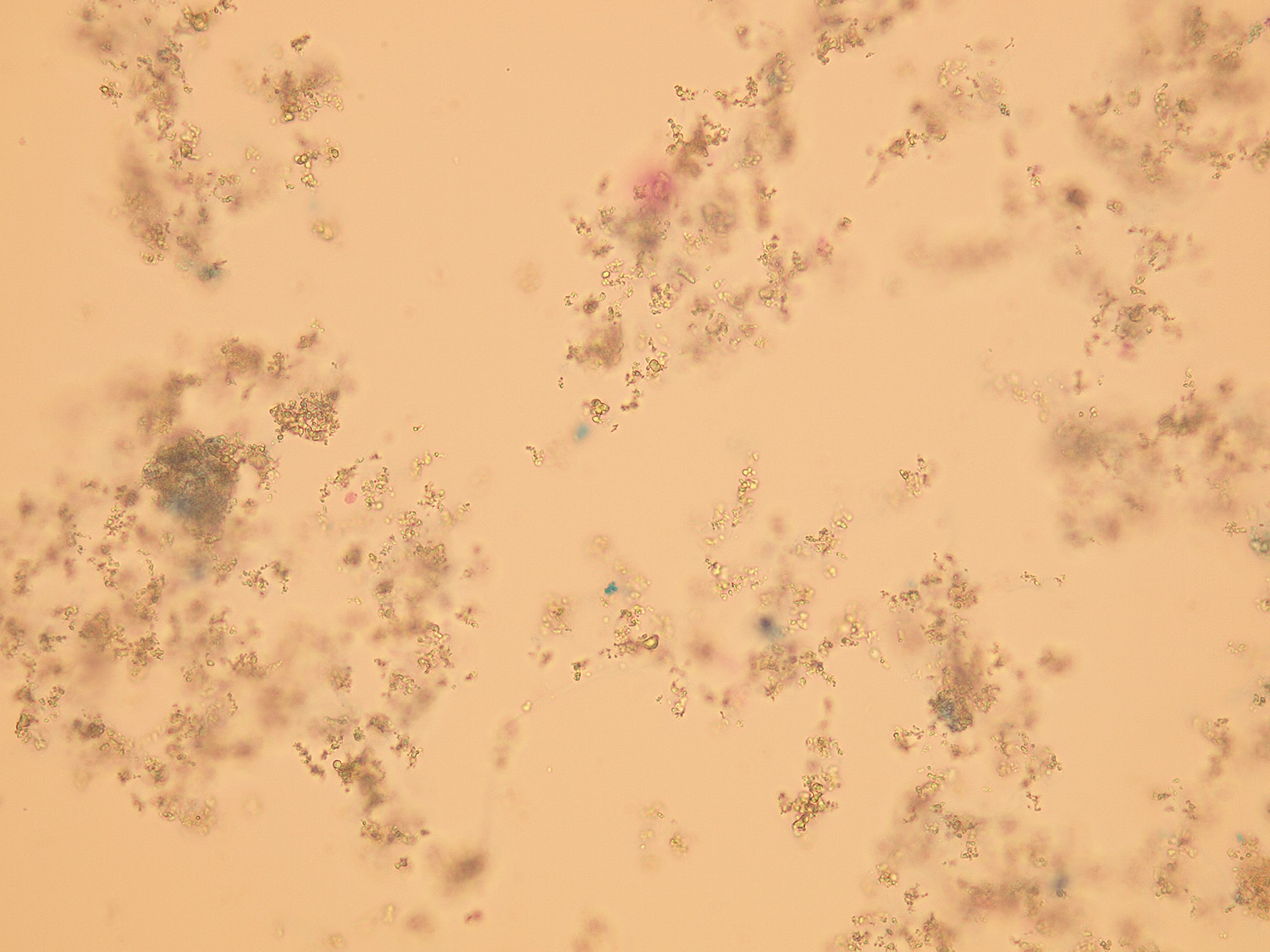
Amorphous microcrystals
Native sediment

Amorphous microcrystals

Amorphous microcrystals
Pictures from iQ 200 analyzer (IRIS)

Amorphous microcrystals

Amorphous microcrystals
Mgr. Ondřej Wiewiorka , MUDr. Jana Tůmová|
KLT, Faculty of Medicine, Masaryk University |
Back to Homepage, accessibility |
| Service Center for E-learning
| Faculty of Informatics, Masaryk University, 2015
Centrum interaktivních a multimediálních studijních opor pro inovaci výuky a efektivní učení | CZ.1.07/2.2.00/28.0041