Size of retinal image calculation in consequences of vertex distance change
Introduction
Size of the retinal image can be changed with shift of the spectacle lens. This way we can decrease value of aniseikonia. In literature we can find limit 3 D which enables image to fuse. Shift with the spectacle lens is recommended if we deal with axial type of aniseikonia.
Goals
- Calculate change in retinal image size during the position change of the minus spectacle lens
- Calculate change in retinal image size during the position change of the plus spectacle lens
Equipment
Minus spectacle lens, plus spectacle lens, calculator, writing equipment
Methods
Calculate change in retinal image size during the position change of the minus spectacle lens
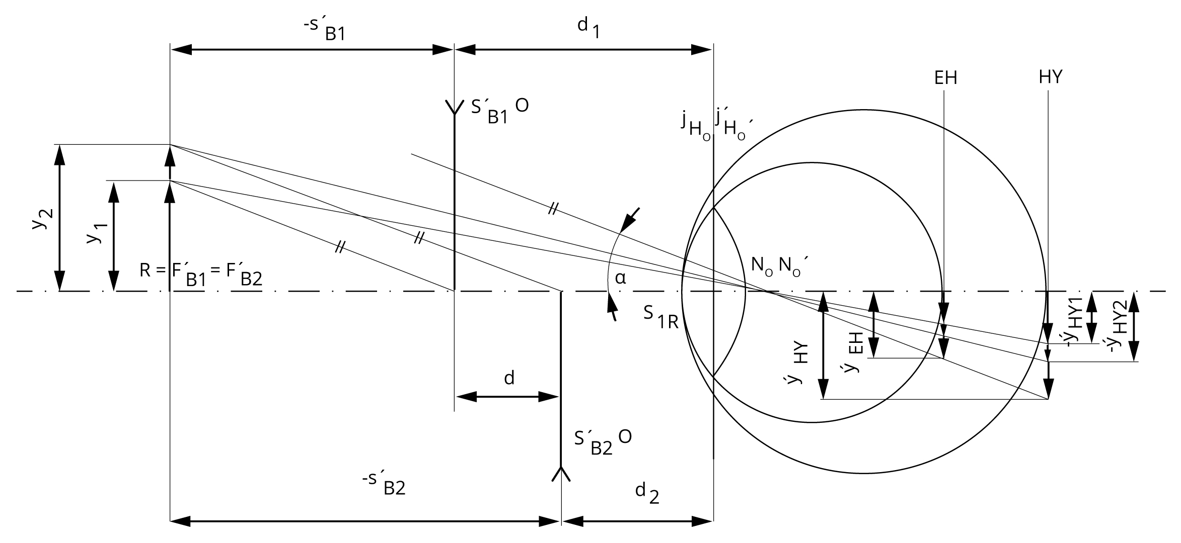
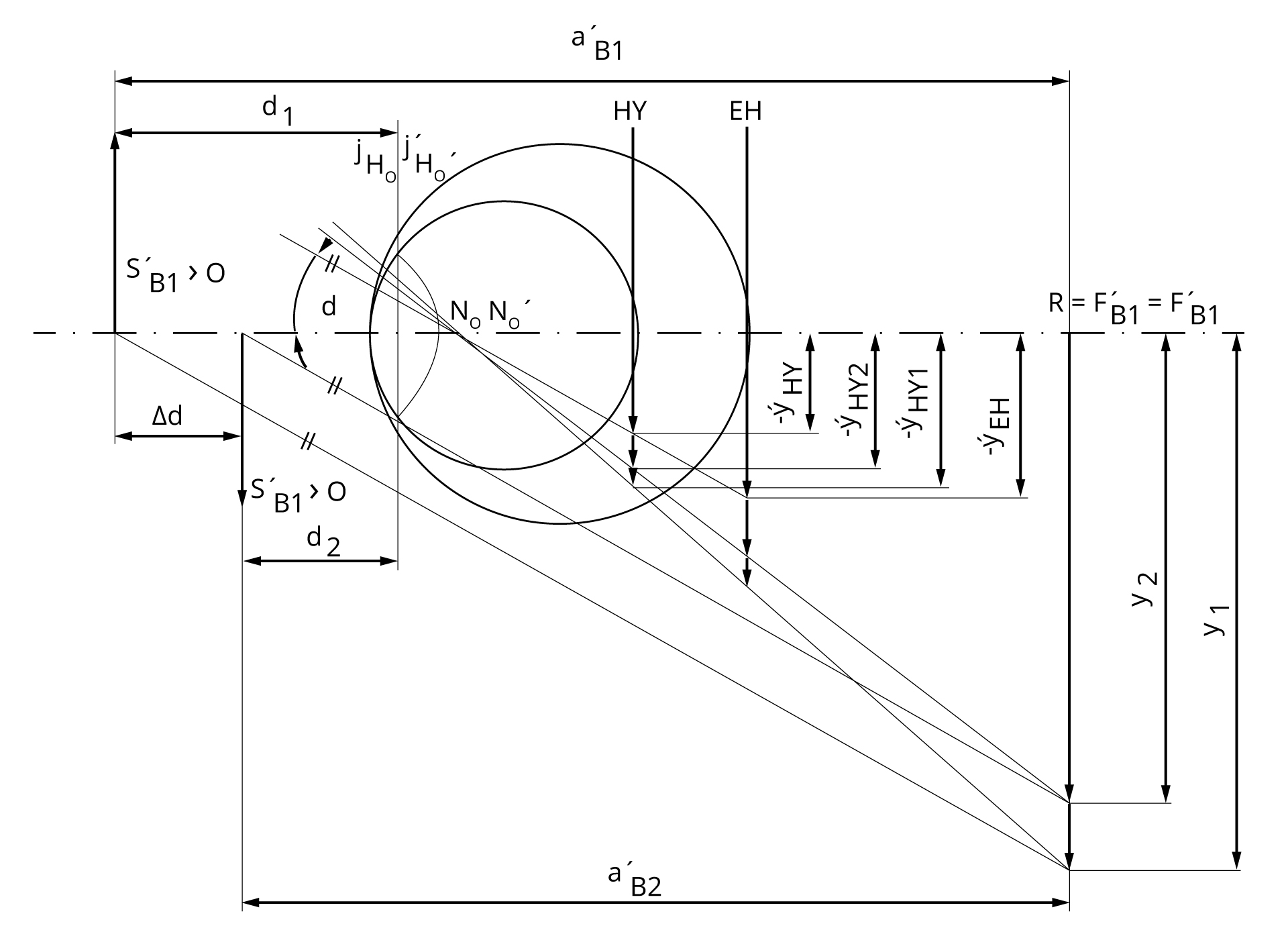
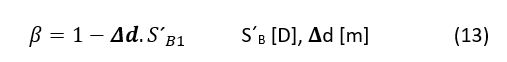
Change of retinal image size depends on spectacle lens position in front of the eye. Ratio between images 𝛃 we can calculate according to ratio between focal distances s´B.
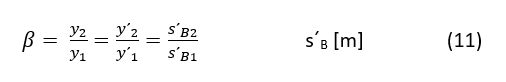
Further according Rutrle (1993) we can conclude
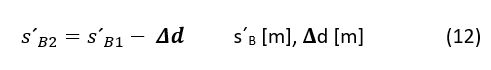
And we substitute from previous formulas
Measure vertex refraction of the minus spectacle lens. We suppose that we change vertex distance with 5 mm to move with the lens closer to cornea. According to above placed formulas calculate change in size of retinal images (𝛃DM)
Calculate change in retinal image size during the position change of the plus spectacle lens
Measure vertex refraction of the plus spectacle lens. We suppose that we change vertex distance with 5 mm to move with the lens close to cornea. According to above placed formulas calculate change in size of retinal images (𝛃DP)
Results
Calculate change in retinal image size during the position change of the minus spectacle lens
𝛃DM =
Calculate change in retinal image size during the position change of the plus spectacle lens
𝛃DP =
Discussion
Generally we can say that in myopia we can find larger image than in hyperopia. In axial hyperopia we usually measure smaller image than in emmetropia. The same size of the retinal image we get if we place corrective lens in eye focus in front of the eye.
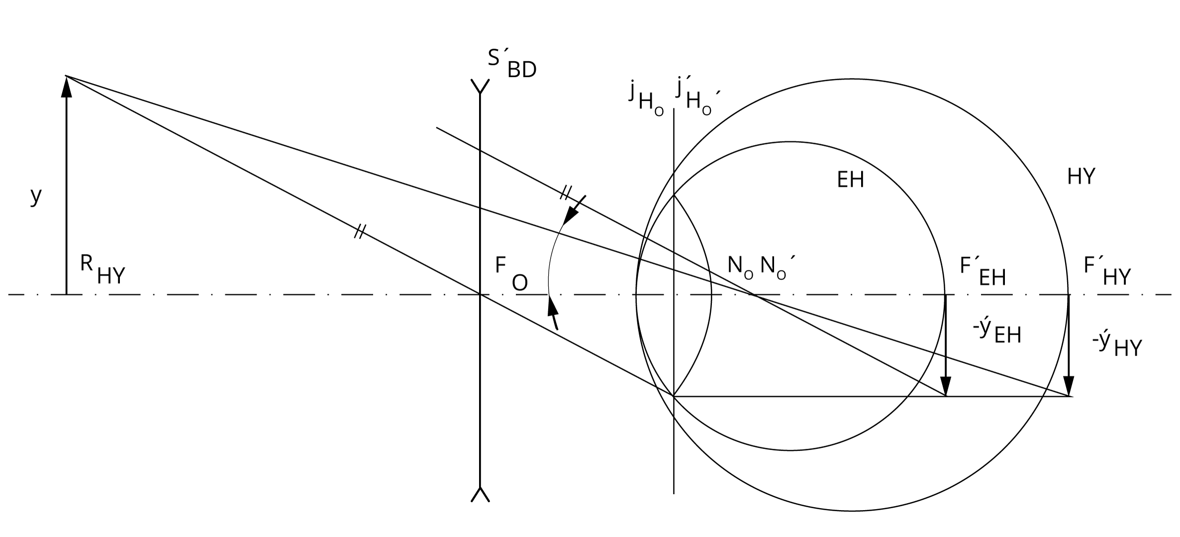
If you shift with minus lens into the eye this increases image before degreasing retinal picture in size. If you shift with plus lens into the eye this degreases image before increasing retinal picture in size.
Conclusion, notes, comments
In which refractive error we can use spectacle lens shit to decrease aniseikonia?
How we can change image size in case of systemic refractive error, i.e. systemic aniseikonia?