Závěrečná práce: Bc. Matyáš Pinkas, učo 451337: Strukturní studie molekulárních strojů in situ
Diplomová práce
Strukturní studie molekulárních strojů in situ
Structural studies of molecular machines in situ
Anotace
Kryogenní elektronova mikroskopie umožňuje studium makromolekulárních komplexů in situ s vyskokým rozlišením za podmínek podobných nativnímu stavu buňky, pomocí technologického postupu kryogenní elektronové tomografie. Celý proces začíná přípravou a vitrifikací buněk, ze kterých jsou nařezány tenké destičky pomocí fokusovaného iontového svazku. Na těchto destičkách jsou poté nasbírána tomografická …více
Abstract
Cryogenic electron microscopy enables high-resolution structural analysis of macromolecular complexes in situ at near-native conditions. This is achieved by utilising cryogenic electron tomography and focused ion beam micromachining. The focused ion beam is applied to produce thin lamellae from vitrified cells and cryogenic electron tomography is employed to acquire tilt-series to compute tomograms …více
Klíčová slova
Skenovací elektronový mikroskop Transmisní elektronový mikroskop Fokusovaný iontový svazek Kryo-elektronová mikroskopie Kryo-elektronová tomografie Ribozom Scanning Electron Microscope Transmission Electron Microscope Focused Ion Beam Cryo-electron Microscopy Cryo-electron Tomography RibosomeZadání práce
Cryo-electron microscopy is a dynamically developing method with increasing utilization in structural biology research. Recent advances in the detector technology enabled near-atomic structural studies of protein complexes including the molecular machines such as RNA polymerases or ribosomes. One advantage of the cryo-electron microscopy with respect to the other high-resolution structural biology methods lies in its capability to structurally characterize pleiomorphic objects and molecular assemblies which are difficult to characterize under in vitro conditions by cryo-electron tomography (cryo-ET). Moreover, when cryo-ET is preceeded by the focused ion beam micromachining (FIBM) of the cellular sample, the resulting cryo-ET tomograms will provide structural information about the macromolecular complexes in the context of the cells at near-native conditions.
The aim of the diploma thesis is to utilize FIBM and cryo-ET to study higher order assemblies of RNA polymerase and ribosome inside Saccharomyces cerevisiae under standard and stress conditions. The student will deepen his knowledge in molecular biology and electron microscopy. She/he will learn to operate both scanning and transmission electron microscopes and analyze the cryo-ET data.
Literature:
- Frank, Joachim, ed. Electron tomography: three-dimensional imaging with the transmission electron microscope. Springer Science and Business Media, 2013.
- Reimer, Ludwig. Scanning electron microscopy: physics of image formation and microanalysis. Vol. 45. Springer, 2013.
- Brandt, Florian, Lars-Anders Carlson, F. Ulrich Hartl, Wolfgang Baumeister, and Kay Grünewald. The three-dimensional organization of polyribosomes in intact human cells. Molecular Cell(2010) 39: 560-569.
- Rigort, Alexander, and Jürgen M. Plitzko. Cryo-focused-ion-beam applications in structural biology. Archives of biochemistry and biophysics (2015), 581: 122-130.
- Rigort, Alexander, Felix JB Bäuerlein, Andrew Leis, Manuela Gruska, Christian Hoffmann, Tim Laugks, Ulrike Böhm et al. Micromachining tools and correlative approaches for cellular cryo-electron tomography. Journal of Structural Biology (2010) 172: 169-179.
3. 6. 2020 15:35, doc. Mgr. Karel Kubíček, Ph.D., učo 20563
 angličtina
Oponent
Katedra biofyziky Přírodovědecké fakulty UP Olomouc
Konzultant
Práce na příbuzné téma
Seznam prací, které mají shodná klíčová slova.
-
Příprava, měření a určování struktur biomolekul metodami elektronové mikroskopie
Mgr. Martina Zánová -
Strukturní a temporální analýza replikace fágů v biofilmu
Ing. Lucie Valentová, Ph.D., učo 436946 -
Porovnání informačního obsahu v různých typech tomografických dat v kryo-elektronové mikroskopii
Mgr. Anna Kasáková -
Strukturní a funkční studie stafylokokového fága phi812
Mgr. Barbora Popelářová -
Cryo-electron microscopy analysis of bacteriophage architecture and infection
Mgr. Ján Bíňovský, Ph.D. -
Materiálový průzkum artefaktů Benátské bible – studium vlivu umělého stárnutí na pigmenty a papírovou podložku a komplexní restaurování dostupných artefaktů
Mgr. Veronika Slachová -
Rozšíření výuky předmětu Struktura a dynamika nukleových kyselin
RNDr. Mgr. Pavlína Pokorná, Ph.D. -
Strukturní charakterizace fosforylované tyrozin hydroxylázy v komplexu se 14-3-3 proteinem
Mgr. Katarína Červená, učo 461069
Složky
Soubory
-
Přidání souboru
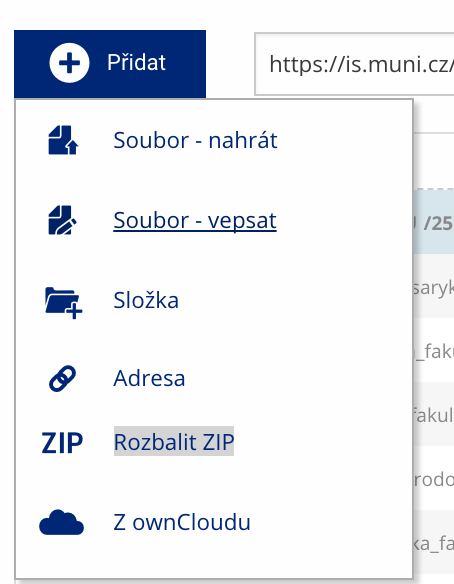 Soubor nebo složku lze nahrát pomocí tlačítka Přidat.
 -
Další operace se soubory
 Podrobnosti lze zjistit označením příslušného řádku.
 -
Pohled pro experty
 Pro častou práci je možné zvolit režim Více možností.
 -
Vyhledávání souborů
 Vyhledávaný výraz můžete zadat přímo do adresního řádku.
 -
Rychlý přístup k souborům
 Pomocí funkce Nedávné je možné se rychle vrátit k právě prohlíženým souborům. Oblíbené soubory je také možné označit Hvězdičkou.